Tag: Particle Size Analysis

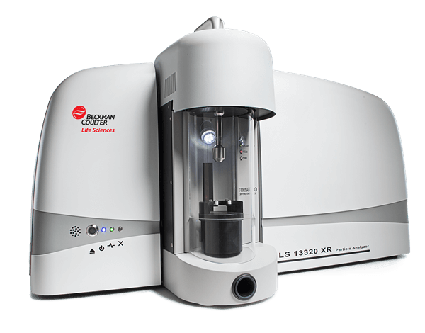
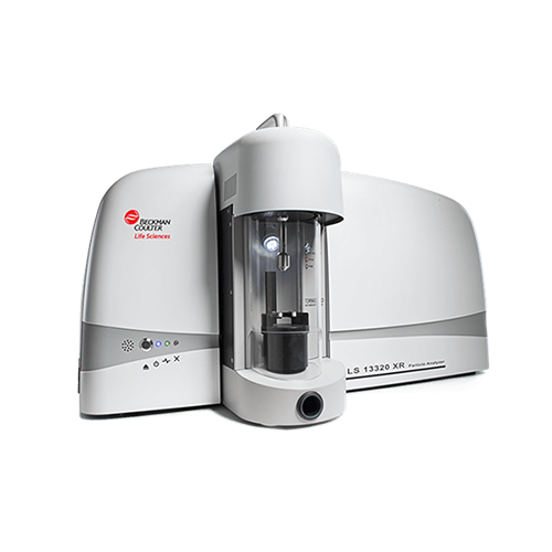
LS 13 320 XR

Beckman Coulter
LS 13 320 XR

Laser Diffraction Particle Size Analyser 

  • Expanded measurement range 10 nm – 3,500 µm
  • Enhanced PIDS Technology
  • Real data down to 10nm

Download
brochure
   Request
quote

Beckman Coulter LS 13 320 XR Laser Diffraction Particle Size Analyser 21 CFR Part 11

Beckman Coulter LS 13 320 XR Particle Size Analyser

For big improvements that help you spot small differences.

The LS 13 320 XR Particle Size Analyser offers best-in-class particle size distribution data from advanced PIDS technology,* which enables high-resolution measurements and an expanded dynamic range. Like the LS 13 320, the XR particle size analyser provides fast, accurate results, and helps you streamline workflows to optimize efficiency. Some big improvements help you reliably spot small differences that can have a huge impact on your particle analysis data.

  • Direct measurement range from 10 nm – 3,500 µm
  • Automatically highlights pass/fail results for faster quality control
  • Enhanced software that simplifies method creation for standardised measurements
  • New control standards to adequately verify instrument/module performance

The LS 13 320 XR Particle Size Analyser is an easy-to-use laser diffraction analyser that yields fast, reliable particle size analysis data for dry and aqueous and non-aqueous samples.

  • Key Features

    Spot Small Differences

    • Particle Size Analyser with expanded measurement range: 10 nm – 3,500 µm
    • Laser diffraction plus advanced Polarization Intensity Differential Scattering (PIDS) technology enable high-resolution measurement & reporting of real data down to 10 nm
    • Provides accurate, reliable detection of multiple particle sizes in a single sample
    Easy-to-use Software

    • ADAPT Software features automatic pass/fail check
    • Pre-configured methods deliver results with 3 clicks or less
    • Simplifies analyser operation by experts & novice users alike
    • 1-click overlay with historical data
    • Intuitive user diagnostics keep you informed during sampling
    • Simplified method creation for standardised measurements
    ADAPT Software enables 21 CFR Part 11
    • Customisable security system to meet diverse needs
    • Choose from 4 security levels
    • High-security configuration supports 21 CFR Part 11
    PIDS Technology* for Direct Detection of 10 nm Particles
    • 3 light wavelengths (450, 600, & 900 nm) irradiate samples with vertical & horizontal polarized light
    • Analyser measures scattered light from samples over a range of angles
    • Differences between horizontally & vertically radiated light for each wavelength yield high-resolution particle size distribution data
  • Technical Specs

    Technology

    Low-angle forward light scattering with additional PIDS(Polarization Intensity Differential Scattering) Technology. Analysis of vertical and horizontal polarized light at six different angles using three additional wavelengths. Full implementation of both Fraunhofer and Mie Theories.

    Light Source

    Diffraction: Laser Diode (785 nm)
    PIDS: Tungsten lamp with high-quality band-pass filters (475, 613 and 900 nm)

    Particle size analysis range

    Measurement range: 10 nm – 3,500 µm
    Dry Powder System Module (DPS): 400 nm – 2,000 µm
    Universal Liquid Module (ULM): 10 nm – 2,ooo µm

    Electrical interface

    USB

    Power consumption

    ≤ 6 amps @ 90 – 125 VAC
    ≤ 3 amps @ 220 – 240 VAC

    Temperature range

    10 – 40°C (50 – 104°F)

    Humidity

    0 – 90% without condensation

    Compliance

    Creates 21 CFR Part 11 enabling features
    RoHS
    Certifications:
    – EU EMC Directive 2014/30/EU
    – CISPR 11:2009/A1:2010
    – Australia and New Zealand RCM Mark

    Data export file formats

    XLSX, TSV, PDF

    File import capability

    From all LS 13 320 Legacy and LS 13 320 XR system

    *Software operating system

    Requires Microsoft Windows 10, 64-bit environment
    (US, English regional settings only)

    Dimensions

    Height: 19.5″ (49.53 cm)
    Width: 37″ (93.98 cm)
    Depth: 10″ (25.4 cm)

    Weight

    52 lbs (23.5 kg)

  • Accessories

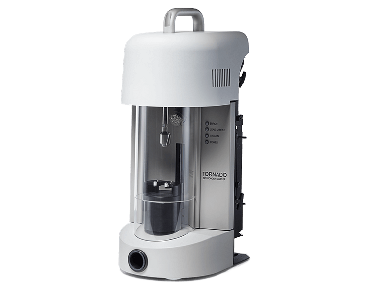
    Dry Powder System

    Analytical size range: 400 nm – 3,500 µm

    • Measures entire sample as required by the ISO 13 320 Standard
    • Programmable Obscuration setting to optimize sample feed rate
    • User-selectable vacuum pressure for maximum dispersion control
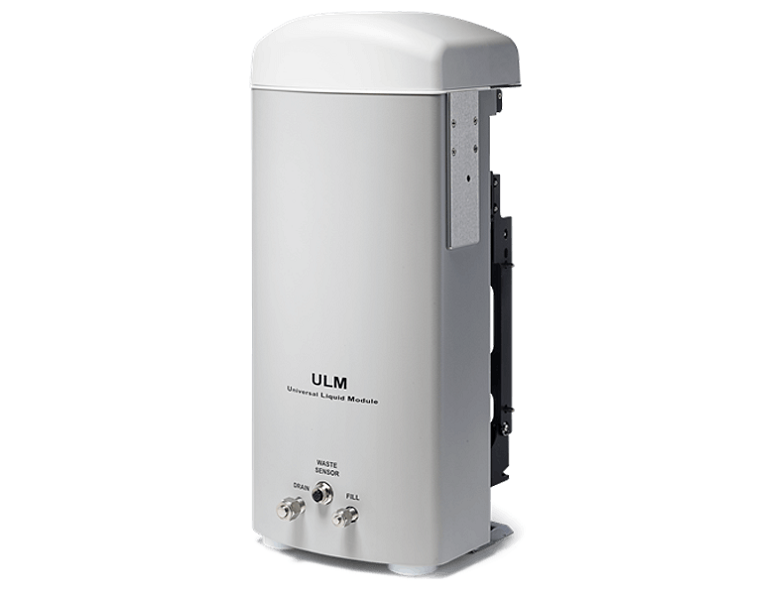
    Universal Liquid Module

    Analytical size range: 10 nm – 2,000 µm

    • Fully automatic with auto-dilution, auto-filling and auto-rinsing
    • Analyses samples suspended in aqueous as well as non-aqueous diluents for maximum flexibility
    • Wetted materials list: Teflon®, 316 Stainless Steel, Glass, Kal-rez® and PEEK
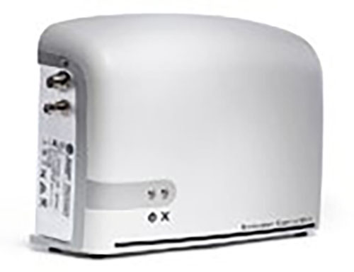
    Sonicator Control Unit
    • Needle probe sonicator for additional dispersion control of wet samples
    • Fully adjustable power settings
    • In-situ sonication with ULM before/during the run, can also be operated external to module

    EU Vacuum Cleaner
    • Vacuum pressure range fully adjustable
    • Integrated vacuum control unit for optimised vacuum/obscuration settings
    • Two vacuum systems to choose from
  • Applications

    Soils

    Nanocellulose

    Pigments

    Cement

  • Capabilities

    The LS 13 320 XR particle size analyzer uses advanced laser diffraction and PIDS technology for the sizing of non-spherical, sub-micron particles. Initially, particle sizing by laser diffraction was limited to the use of the Fraunhofer diffraction theory. Laser diffraction offers a number of advantages – laser diffraction analyzers go beyond simple diffraction effects. General approaches are now based on the Mie theory and the measurement of scattering intensity over a wide scattering angular range is employed.

    Using PIDS Technology

    Pioneered by Beckman Coulter, most laser diffraction manufacturers use the above two approaches, i.e., wide angular detecting range and short wavelength, to size small particles. However, sizing even smaller particles (tens of nanometers in diameter), cannot be achieved using only these two approaches. Any further increase in scattering angle will not yield any significant improvement due to the everslower angular variation. Figure 2 is a 3-D display that illustrates the very slow angular variation for small particles. For particles smaller than 200 nm, even by taking advantage of the above two approaches, it is still difficult to obtain an accurate size.
Not sure if it’s the right instrument?

No worries, send us a sample and we will test it for you


Send a sample

Continue reading

Multisizer 4e

Beckman Coulter
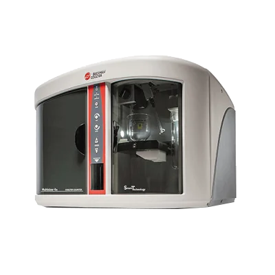
Multisizer 4e

Coulter Coulter – Particle Size and Count Analyser

  • Unparalleled sizing range of 0.2 – 1600 μm
  • Advanced noise reduction system
  • Patented digital pulse processing technology

Download
brochure
   Request
quote

The Multisizer 4e provides accurate particle and cell counting. It is the most accurate and flexible particle characterisation device available, boasting an unparalleled sizing range of 0.2 – 1600 μm. The new 10 μm Aperture allows users to study sub-cellular and micro-particles as small as 200 nm, while the advanced noise reduction system for small apertures improves measurement accuracy.

Accurate particle and cell counting

The Multisizer 4e Coulter Counter is used in a number of different fields for counting and sizing both particles and cells. Applications include: Quality Control, Research and Development, Pharmaceutical Analysis, Biotechnology Applications and Industrial Applications.

  • Key Features

    Versatility
    • Aperture sizes from 10 µm – 2000 µm in diameter for current & future needs
    • Multiple aperture options & data overlay capability for analyzing complex samples over wide particle size distribution
    Sample Detection
    • Analyse materials in aqueous or non-aqueous solvents
    • Ideal for biological or non-biological samples
    • Detect particles regardless of material type or optical properties
    Rapid Particle Counting
    • Typical sampling rates up to 10,000 counts per second
    • Detect & count particles from 0.2 µm – 1600 µm in diameter
    • Sample volumes as small as 5 mL
    Data Management
    • Digital pulse data can be stored & re-analysed as needed
    • CFR 21 Part 11 compliant software for audit control & security
    • Variety of calibration beads & V-check validation controls
  • Technical Specs

    Particle Sizing Range

    Diameter: 0.2 – 1,600 µm
    Volume: 0.004 – 2.145 x 109 µm3

    Aperture Size

    10 – 2,000 µm (nominal diameter)

    Measurement range

    Extended: 2 – 80% of aperture size
    Standard: 2 – 60% aperture size

    Measurement linearity

    Diameter: ± 1%
    Volume: ± 3%

    Dynamic range (accuracy)

    Diameter: 1 : 40 (extended), 1 :30 (standard),
    Volume: 1 : 64,000 (extended), 1: 27,000 (standard)

    Processor type

    High speed signal digitalisation

    Number of pulses measured

    Up to 525,000 per analysis

    Resolution

    User difined

    Number of size classes

    Up to 400 for display of any selected measurement range
    The number of classes and their width can be changed as needed

    Pulse distribution data

    X axis: time, registration sequence, pulse width
    Y axis: diameter, volume or voltage corresopnding to pulse amplitude,
    pulse width, mean diameter, volume, or voltage corresponding to pulse
    amplitude, average pulse width, pulse distribution by width

    Particle size distribution data

    X axis: diameter, volume, surface area
    Y axis: absolute number, percent content (%), number per ml, absolute volume, volume percentage (%) volume per ml, absolute surface area (%) surface area per ml

    Sample registration mode:
    total number of particles

    50 – 500,000 counts

    Sample registration mode:
    number of particles and
    measurement of parameters

    10 – 100,000 counts

    Sample registration mode:
    time

    0.1 – 999 seconds with 10 ms increments
    Standard registration time is 10 – 90 seconds

    Sample registration mode:
    volume

    50 – 2,000 µL

    Dosage system

    The dosing pump with even suspension flow across the aperture and volume measurement, error – less than 0.5%

    Electrolyte type:

    Aqueous and non-aqueous electrolyte solutions compatible with glass, fluoropolymers, fluoroelastomers and stainless steel

    Aperture current strength range:

    30 – 6,000 µA with 0.2µA increments

    Aperture current stability:

    ± 0.4% of set value

    Polarity error

    Less than 0.5%

    Compliance with standards

    Software is 21 CFR part 11 compliant

    Dimensions

    64 x 61 x 51 cm, weight 45 kg

    Power supply requirements

    230 – 240 V ± 10%, 47 – 63 Hz

    Power consumption

    Less than 55 Watts

    Fuses

    250 V, IEC (5×20 mm) with time delay, 2.0 A

    Environmental requirements

    The instrument is intended for work in enclosed spaces
    Working temperature: 5 – 40°C
    Relative humidity: 30 – 85% without condensation

  • Applications

    Yeast

    Beer

    Protein

    E. coli

    Plant Cells

    CAR-T Cells

  • Capabilities

    The Multisizer 4e particle sizer and counter is the most accurate and flexible particle characterization device available, boasting an unparalleled sizing range of 0.2 – 1600 μm. The new 10 μm Aperture allows users to study sub-cellular and micro-particles as small as 200 nm, while the advanced noise reduction system for small apertures improves measurement accuracy.

    Generated data are processed using patented digital pulse processing technology and can be saved and later re-analyzed. This technology provides ultra high resolution and accuracy unattainable through any other technologies: detection of 1 particle in 1 ml of a sample with the optimal instrument configuration. Analysis results are not dependent on particle shape, structure, or optical properties.

    It uses the Coulter principle to detect particles via electrical zone sensing, regardless of the particle’s nature or optical properties. This makes it an ideal tool for detecting and counting a wide variety of particles, such as:

    • Mammalian cells
    • Bacteria
    • Yeast
    • Abrasives
    • Toner particles
    • Cell aggregates
    • Spheroids
    • Large protein aggregates

Not sure if it’s the right instrument?

No worries, send us a sample and we will test it for you


Send a sample

Continue reading

Bettersizer S3 Plus

Bettersize
Bettersizer S3 Plus

Particle Size and Shape Analyser 

  • 0.01 – 3,500 µm (laser system)
  • 2 – 3,500 µm (image system)
  • 0.5x and 10x cameras
  • Rapidly generates results in 10 seconds

Download
brochure
   Request
quote

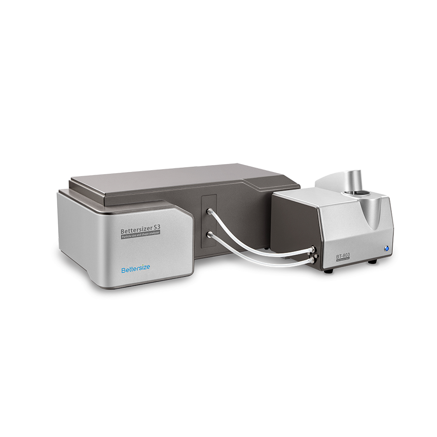
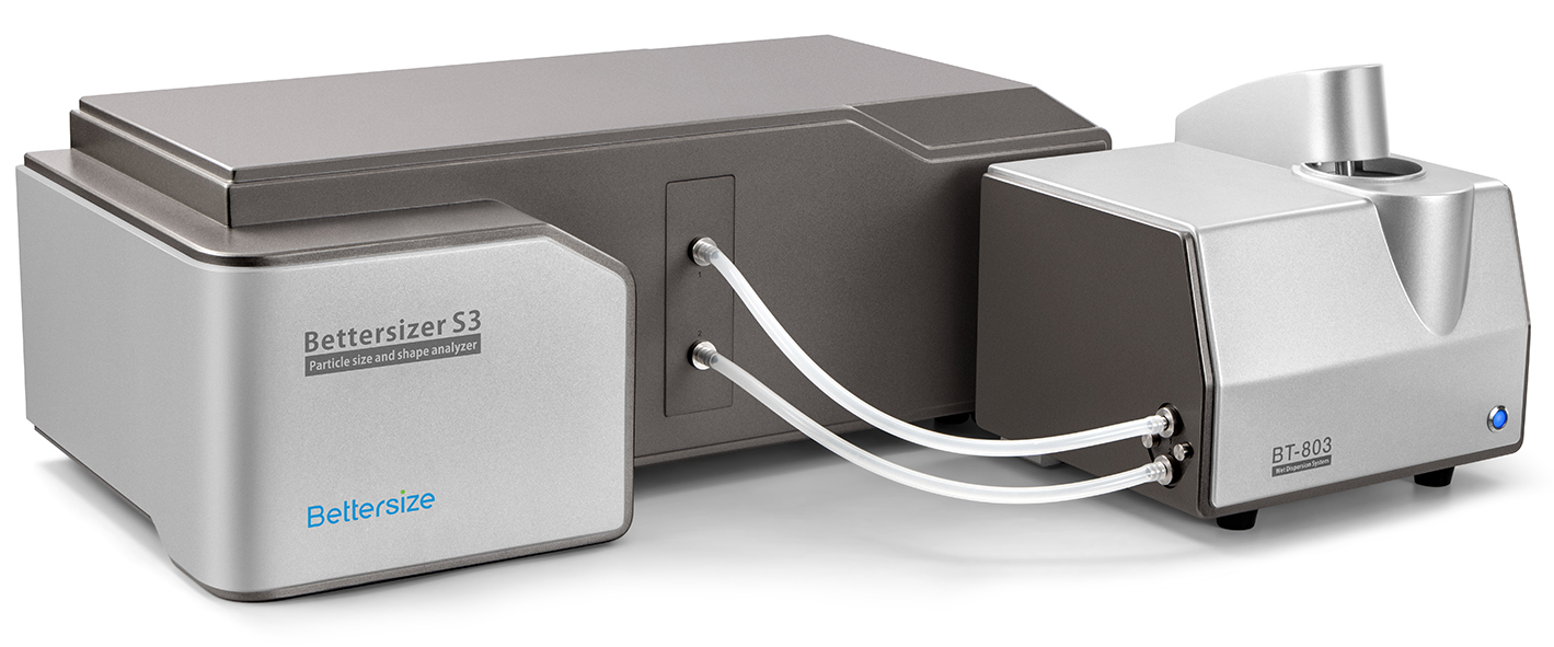
Bettersizer S3 Plus+BT-803

Strive for excellence in all you see.
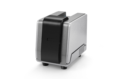
The Bettersizer S3 Plus particle size and shape analyser combines laser diffraction and dynamic image analysis in one instrument. It can measure the size and shape of particles from 0.01 µm to 3500 µm. Its exceptional sensitivity for either ultrafine particles or oversized particles, and unsurpassed resolution, make it the most powerful size and shape analyser for enthusiastic researchers who conduct top scientific research.
  • Features and Benefits

    • Measuring range is 0.01 – 3,500µm (laser system), 2 – 3,500µm (image system)
    • Combining laser diffraction and dynamic image analysis in one instrument, obtaining size and shape results simultaneously
    • Patented DLOI (Dual Lenses & Oblique Incidence) system enable the measurement of ultrafine particles down to 0.01 µm
    • Dual-camera imaging technology can show particles images in real time and detect oversized particles up to 3500 µm
    • Refractive index measurement determines the refractive index of unknown samples and improves te reliability of results
    • Compliance with 21 CFR Part 11, ISO 13320, USP <429>, CE
  • Performance

    1) High Resolution and Sensitivity

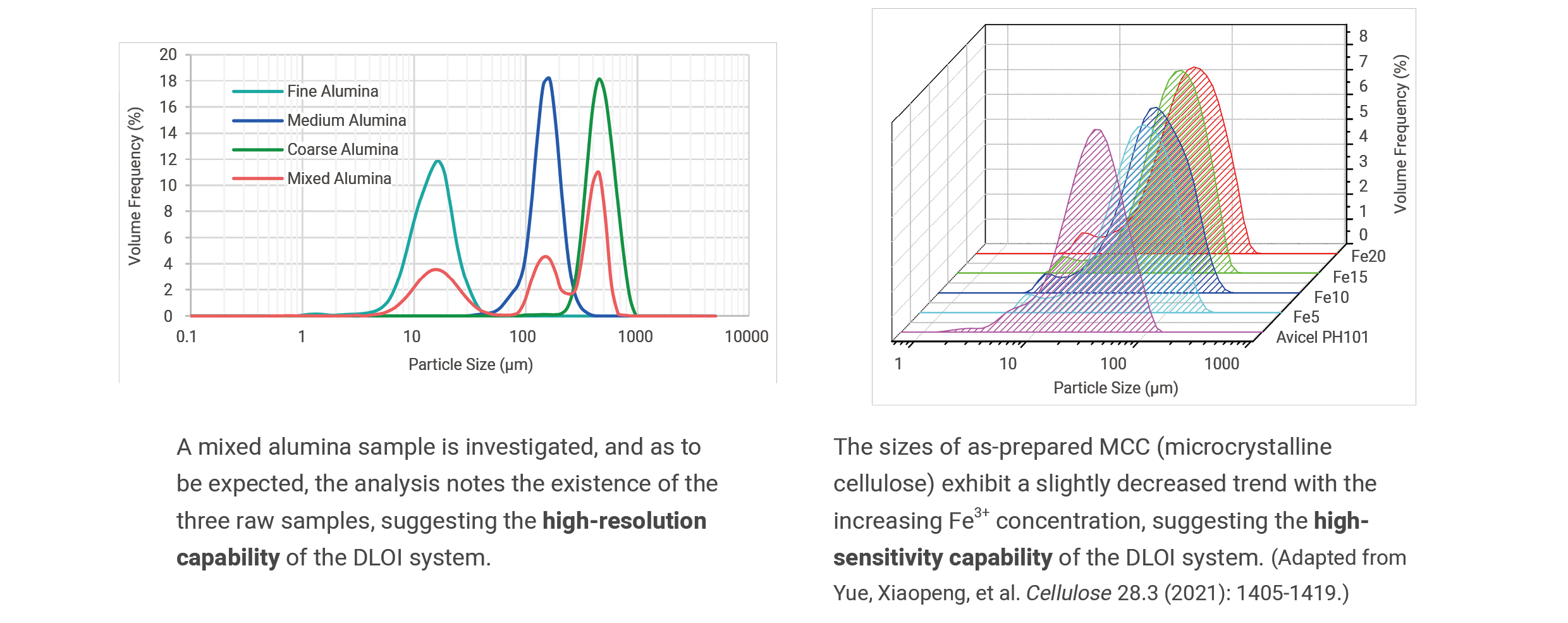
    The Bettersizer S3 Plus achieves exceptional resolution and sensitivity for particle size measurements. The DLOI system allows the size distributions of polydisperse samples to be determined precisely, and the size changes of products to be detected sensitively.

    BioLector Microbioreactor

    2) Multiple Shape Parameters

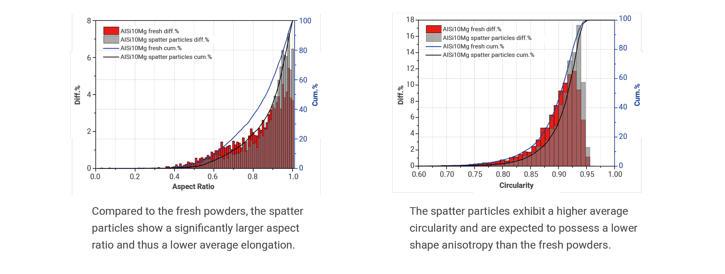
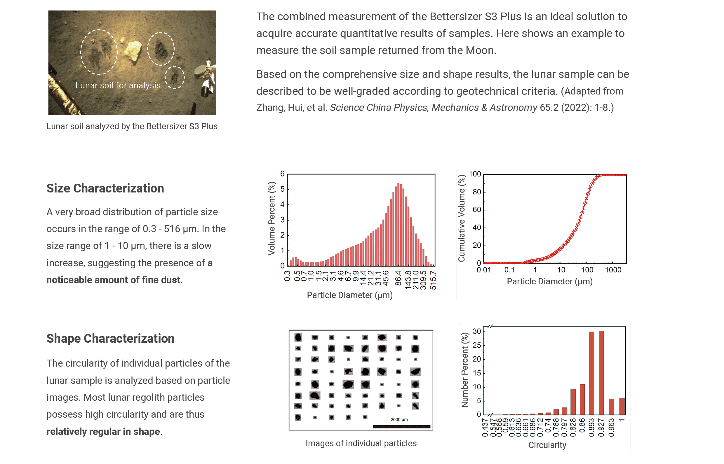
    An example of additive manufacturing for shape analysis using the Bettersizer S3 Plus is shown below. A representative number of individual particles are recorded from two AlSi10Mg samples, and the number-weighted aspect ratio and circularity are evaluated in compliance with ISO standards. (Adapted from F. Schleife, C. Oetzel. Chem. Ing. Tech. 93.8 (2021): 1199–1203.)

    BioLector Microbioreactor

    3) Oversized Particle Detection

    Laser diffraction in combination with image analysis can sensitively detect oversized particles that are statistically underrepresented within a wide-distributed sample, such as oversized grains, agglomerates, air bubbles, etc. An example of an off-specification abrasive is displayed below. The Bettersizer S3 Plus confirms the presence of oversized particles, by showing a size peak at around 120 μm and the images of overly coarse particles.

    BioLector Microbioreactor

    4) Analysis of Samples with Extremely Broad Distributions

    BioLector Microbioreactor

  • Technology

    Image Analysis

    Laser Diffraction

  • Applications

    Soy Milk

    Advanced Ceramic Products

    Powder Coatings

    Ceramic Agglomerates

    Lithium-Ion Batteries

    Soils and Sediments

    Abrasives

    3D Printing

    Differing Abrasives

  • Specification

    Particle size distribution

    Suspension, emulsion, dry powders

    Particle shape

    Suspension, emulsion, dry powders

    Principle

    Laser diffraction and dynamic image technologies

    Analysis

    Mie scattering theory and Fraunhofer diffraction theory

    Typical measurement time

    Less than 10 seconds

    Measuring range

    0.01 – 3500 μm (Laser System)
    2 – 3500 μm (Image System)

    Accuracy

    <0.5% (NIST certified standards)

    Repeatability

    <0.5% (NIST certified standards)

    Number of size classes
    ≤100 (adjustable)
    Feeding mode

    Automatic circulation or semi-automatic circulation

    Special functions

    Refractive index measurement, SOP settings

    Image recognition

    Up to 120 fps, up to 10,000 particles per min

    Optical system

    Patented DLOI (Dual Lenses & Oblique Incidence) System

    Laser

    Polarized light-pumped solid-state laser (10 mW / 532 nm)

    Detector

    96 detectors (forward, lateral and backward arrangements)

    Measuring angle

    0.02 – 165°

    CDC cameras

    0.5x and 10x *

    Image analysis

    1.2 megapixels

    Circulation speed

    300 – 2500 r/min 

    Circulation flow rate

    3000 – 8000 mL/min 

    Ultrasonication

    Dry run protection, Max 50 W (adjustable) 

    Circulation tank capacity

    600 mL

    Conformity

    21 CFR Part 11, ISO 13320, ISO 13322, USP <429>, CE 

    Report

    Customizable reporting

    Dimensions (L x W x H)

    820 × 610 × 290 mm 

    Weight

    48 kg

    Voltage

    DC 24 V, 50 / 60 Hz, 20 W

    Computer interface

    At least one high-speed USB 2.0 or USB 3.0 port required

    Operating system

    Windows 7 / Windows 10

    Hardware specification

    Intel Core i7, 8GB RAM, 500GB HD, two PCI-E X16 interfaces

    * The Bettersizer S3 Plus is also available in a single camera (0.5x) model. Contact us for more information.

  • Accessories

    Introduction

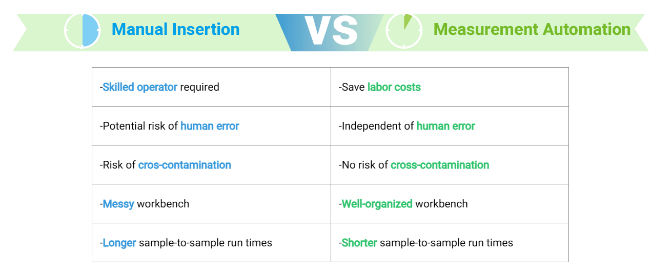
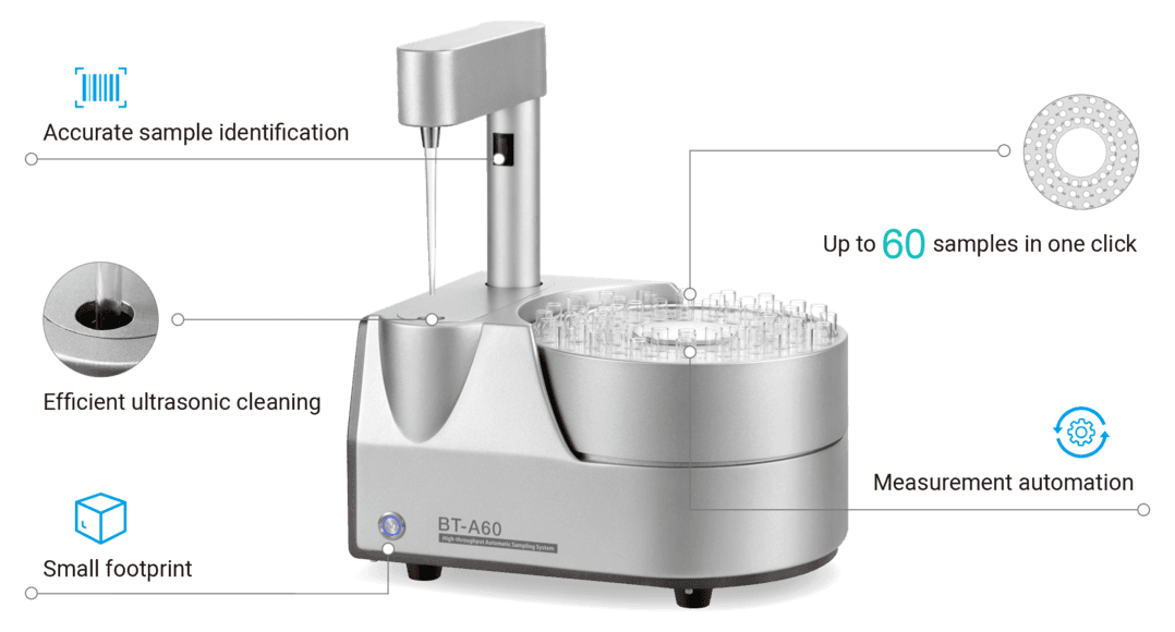
    The BT-A60 is a durable, automatic and high-throughput sampling system. It delivers maximum laboratory automation for sample measurements, reducing your labor costs while improving productivity and laboratory efficiency. The compact design saves valuable bench space while allowing up to 60 different samples to be measured in a single run. Compatible with Bettersizer S3 Plus and Bettersizer 2600, the BT-A60 offers 24/7 fully automated sample analysis to meet your various analytical applications.

    Features

    • Accurate sample identification
    • Efficient ultrasonic cleaning
    • Up to 60 samples in one click
    • Measurement automation
    • Small footprint
    BioLector Microbioreactor

Not sure if it’s the right instrument?

No worries, send us a sample and we will test it for you


Send a sample

Continue reading

Bettersizer 2600

Bettersize
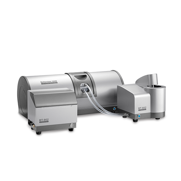
Bettersizer 2600

Laser Diffraction and Dynamic Image Analysis

  • Wet dispersion: 0.02 to 2,600µm
  • Dry dispersion: 0.1 to 2,600µm
  • Dynamic Imaging 2.0 to 3,500µm 

Download
brochure
   Request
quote

Bettersizer 2600 Particle size Dynamic image analysis Laser diffraction

The Bettersizer 2600 utilises proven Laser Diffraction Technology to measure particle sizes ranging from 0.02 to 2,600 μm. Its modular design provides versatile functionality, with the dynamic imaging module enabling combined laser and imaging tests. This extends the measurement range to 3,500 μm while allowing for both particle size and shape analysis. Additionally, the system’s flexible dispersion modules support both dry and wet dispersion methods, catering to a wide range of measurement needs.

  • Key Features

    Technology

    Laser Diffraction

    Particle size range

    Wet dispersion: 0.02 to 2,600 µm Dry dispersion: 0.1 to 2,600 µm

    Sample state

    Wet and dry method of particle sizing is suitable for a wide variety of dispersing particle systems

    Range

    Precise measurement of both large and small particles covering the broadest range by a new patented technology utilizing both Fourier and Reverse Fourier optical systems

    Detectors

    Optical bench with 92 detectors covering an angular range of 0.016° – 165° Optional wet and/or dry dispersion

    Small Volume

    Small volume dry dispersing module for small sample quantities especially for pharmaceutical or valuable samples in short supply

    Speed

    Easy and fast to changeover between dispersing modules

    SOP’s

    Easy to implement, create and use Standard Operation Procedures for new materials

    Refractive Index Measurement

    Refractive index measurement for providing a more accurate parameter to calculate the results.

    Software
  • Specification

    Particle size distribution

    Suspensions, emulsions, dry powders

    Principle

    Laser diffraction technology

    Analysis

    Mie scattering theory and Fraunhofer diffraction theory

    Measurement time

    Less than 10 seconds

    Measuring range

    Wet dispersion: 0.02 to 2,600 µm Dry dispersion: 0.1 to 2,600 µm

    Accuracy error

    ≤ 0.5% *

    Repeatability

    ≤ 0.5% *

    Number of size classes

    100 (user adjustable)

    Feeding mode

    Automatic circulation or micro cuvette (wet)

    Gas transportation (dry)

    Special functions

    SOP settings, refractive index measurement, sample ratio calculation)

    Optical System

    Combined Fourier and inverse Fourier & Tilted sample cell

    Laser

    High-power optical fiber laser (10 mW / 635 nm)

    Detector

    92 detectors (forward, lateral and backward arrangements)

    Measuring Angle

    0.016 – 165°

    Measurement method

    One-click operation (automated measurement, cleaning, saving, printing, etc.)

    Dispersion medium

    Water or organic solvents (sample dependent)

    Circulation

    300 – 2500 r/min

    Ultrasonication

    Dry burning prevention,50 W

    Dispersion medium

    Air/ Nitrogen/Noble gas

    Air pressure

    0.1 – 0.8 MPa (Air compressor dependent)

    Conformity

    21 CFR Part 11, ISO 13320, USP <429>, CE

    Reports

    Customisable reporting

    Dimensions

    70.5 x 31.8 x 29.5 cm

    Weight

    23 kg

    Supply voltage

    100 – 240V, 50/60 Hz

    Weight

    52 lbs (23.5 kg)

    Computer Interface

    At least one high-speed USB 2.0 or USB 3.0 port required

    Operating system

    Windows 7 (32 bit and 64 bit) or higher

    Hardware specification

    Intel Core i5 Processor, 4GB RAM, 250GB HD, Wide screen monitor

  • Accessories

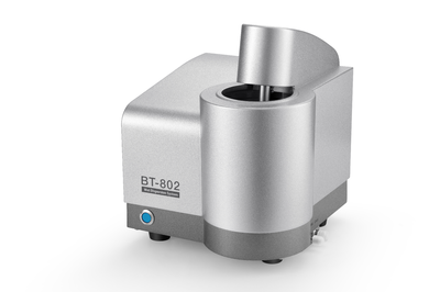
    BT-802

    Wet method

    600mL (dispersion medium)

    Fully automated

    BT-804

    Wet & small volume

    8mL (dispersion medium)

    Semi-automated

    BT-80N

    Anti-corrosive

    80mL (dispersion medium)

    Semi-automated

    BT-902

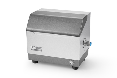
    Dry method

    0.2 – 10 g (sample)

    Fully automated

    BT-903

    Dry & small volume

    0.02 – 1 g (sample)

    Fully automated

  • Applications

    Pigments

    Coffee

    Chocolate

    Ceramic Powders

    Lithium Batteries

    Powder Coatings

    Abrasives

    Mineral Pigment

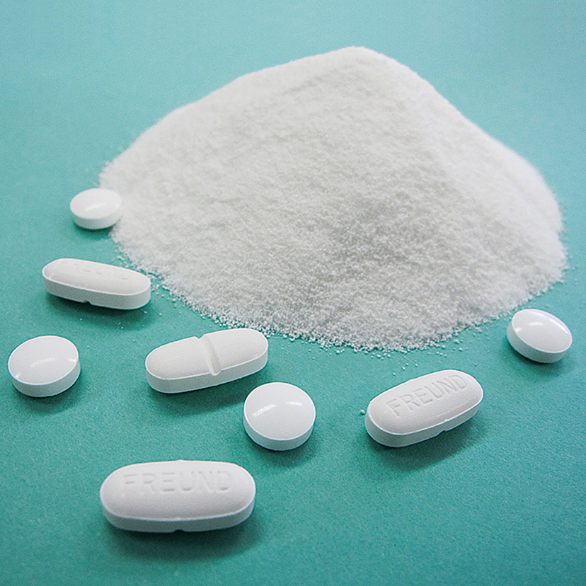
    Pharmaceuticals

    Powdered Milk

    Cement

    Coffee 2

    Lactose

    Pharmaceuticals 2

    Chinese Medicine

  • Technology

    Laser Diffraction

Not sure if it’s the right instrument?

No worries, send us a sample and we will test it for you


Send a sample

Continue reading

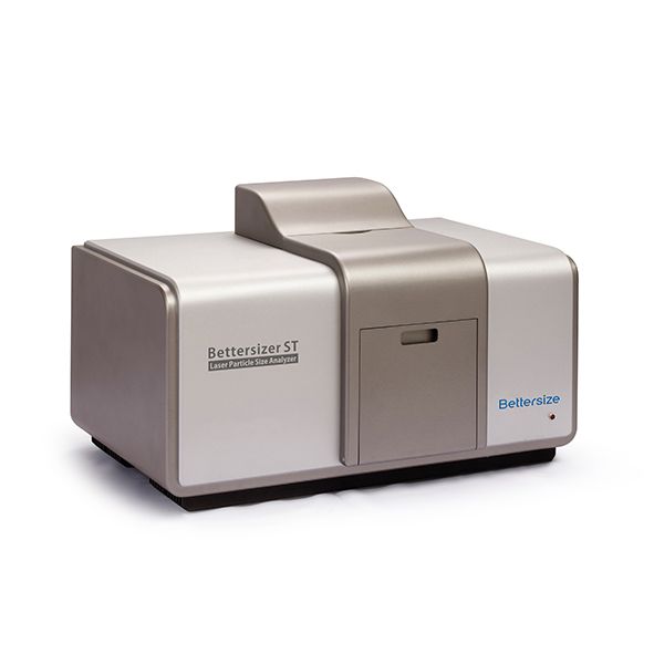
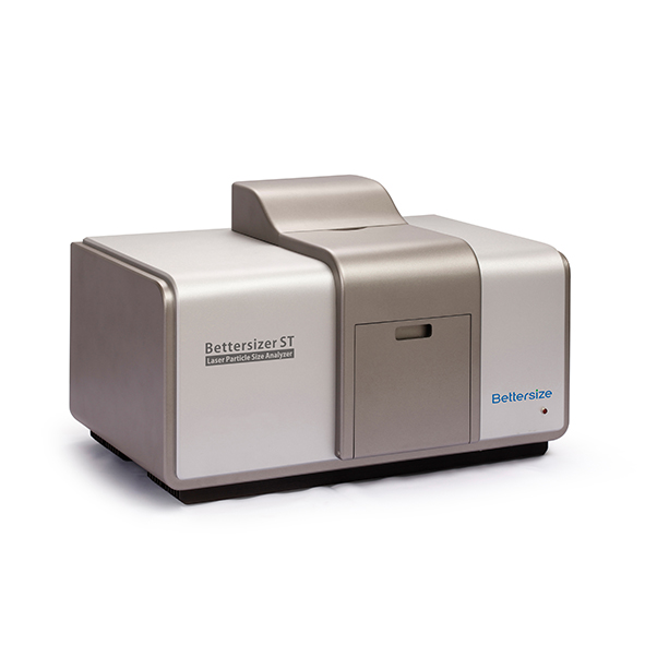
Bettersizer ST

Bettersize
Bettersizer ST

Particle Size Analyser – Wet Analysis

  • Size range 0.1 – 1,000 µm
  • Low maintenance
  • Cost-Efficiency

Download
brochure
   Request
quote

Beckman Coulter LS 13 320 XR Laser Diffraction Particle Size Analyser 21 CFR Part 11

The Bettersizer ST is a fully automated and integrated particle size analyser with a smart operation system by wet dispersing. Optimised for the industrial quality control process, the Bettersizer ST provides stable and reliable testing results with minimum user intervention. The compact footprint saves valuable workspace for factories and laboratories.

Particle Size Analyser you can TRUST

The Bettersize Bettersizer ST Particle Size Analyser ensures reliable wet analysis for quality control processes. Utilising laser diffraction, it provides accurate and reproducible measurements. Ideal for various industries, the Bettersizer ST guarantees precise particle size distribution, enhancing product consistency and quality assurance

  • Key Features

    • Dispersion type: Wet
    • Size range: 0.1 – 1,000µm
    • Robustness
    • Ease-of-Use
    • Cost-Efficiency
    • Compact design
    • Low maintenance
    • Dual lens system
    • Automatic alignment functionality
    • Automatic measurement with SOP
  • Technical Specs

    Particle Size Distribution

    Suspensions, emulsions, dry powders

    LPrinciple

    Laser diffraction technology

    Analysis

    Mie scattering theory and Fraunhofer diffraction theory

    Typical measurement time

    Less than 10 seconds

    Measurement range

    0.1 µm – 1000 µm

    Accuracy error

    ≤1% (NIST certified standards)

    Repeatability error

    ≤1% (NIST certified standards)

    Number of size classes

    ≤100 (adjustable)

    FOptical system

    Patented DLOS (Dual Lens Optical Systems)

    Laser

    High-power fiber semiconductor laser (10 mW/635 nm)

    Detector

    86 photodetectors (forward, lateral and backward arrangements)F

    Measuring angle

    0.031 – 159°

    Circulation speed

    300 – 2500 r/min

    Circulation flow rate

    3,000 – 8,000 mL/min

    Ultrasonication

    Dry run protection, Max 50 W (adjustable)

    Circulation tank capacity

    600 mL

    Conformity

    21 CFR Part 11, ISO 13320, CE

    Reports

    Customizable reporting

    Dimensions (L x W x H)

    660 x 420 x 320 mm

    Weight

    38 KG

    Voltage

    DC 24V, 221 W

    Computer interface

    At least one high-speed USB 2.0 or USB 3.0 port required

    Operating system

    Windows 7 or higher

    Hardware specification

    Intel Core I5, 4GB RAM, 250GB HD

  • Applications

    Lithium-ion Battery

    Calcium Carbonate

    Pesticides

    Ceramic Powder

    Abrasives

    Gypsum

    Pesticides 2

  • Technology

    Laser Diffraction

Not sure if it’s the right instrument?

No worries, send us a sample and we will test it for you


Send a sample

Continue reading

BeNano 180 Zeta Pro

Bettersize
BeNano 180 Zeta Pro

Nanoparticle Size and Zeta Potential Analyser

  • Expanded measurement range 10 nm – 3,500 µm
  • Enhanced PIDS Technology
  • Real data down to 10nm

Download
brochure
   Request
quote

Beckman Coulter LS 13 320 XR Laser Diffraction Particle Size Analyser 21 CFR Part 11

The BeNano Series is the latest generation of nanoparticle size and zeta potential analysers designed by Bettersize Instruments. Dynamic light scattering (DLS), electrophoretic light scattering (ELS), and static light scattering (SLS) are integrated into the system to provide accurate measurements of particle size, zeta potential, and molecular weight. The BeNano Series is widely applied in academic and manufacturing processes of various fields including but not limited to: chemical engineering, pharmaceuticals, food and beverage, inks and pigments, and life science, etc.

Features and Benefits

  • Size range: 0.3nm – 15μm
  • Minimum sample volume 3μL
  • APD (Avalanche Photodiode) detector providing exceptional sensitivity
  • Automatic adjustment of laser intensity
  • Intelligent algorithm of result evaluation
  • DLS backscattering (173°) detection technology
  • User-adjustable scattering volume for concentrated samples
  • PALS (Phase Analysis Light Scattering) technology
  • Programmable temperature control system
  • Compliance with 21 CFR Part 11, ISO 22412, ISO 13099

  • Features

    1) Unlock Greater Research Potential With BeNano

    • Advanced ELS Technology: PALS

      PALS technology can effectively distingguish and extract the electrophoretic behavior even for sample with weak eletrophoretic mobilities, either close to isoelectrical point or with high salinity environment.

    • Advanced DLS Technology: Backscattering Detection

      Backscattering DLS optics can detect much larger scattering volume compared to 90-degree optics. Combined with movable measurement position, backscattering DLS offers much higher sensitivity and high turbidity sample measurement capacity.

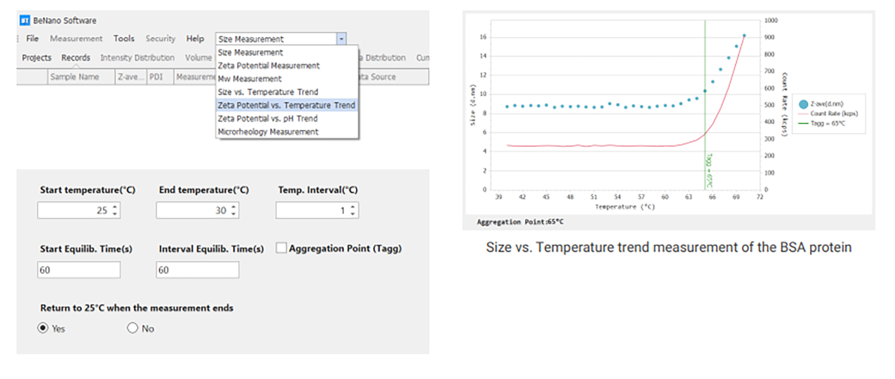
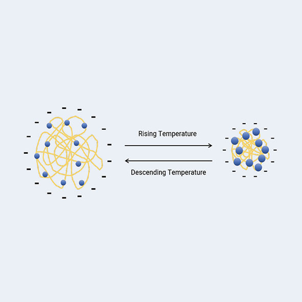
    • Temperature Trend Measurement

      For thermal sensitive samples, a temperature trend can be performed easily with a programmed SOP. The BeNano can detect the temperature transition point of the size results, which is the aggregation temperature for protein samples.

    • Stable and Durable Optical Bench

      The BeNano adopts a 50mW solid-state laser, a singlemode fiber system and a high-performance APD detector, providing stable, wide-ranging, and highly redundant detection capabilities.

    • Research Level Software

      The BeNano software can evaluate and process scattered light signals intelligently to improve the signal quality and result stability. Various built-in calculation modes can cover multiple scientific research and application fields.

    • Trace Sample Volume

      Measuring trace amount of sample is required for earlystage R&D in pharmaceutical industy and academia. With the capillary sizing cell, only 3 to 5 μL of sample is needed for precise size measurement.

    2) Particle Size Measured by Dynamic Light Scattering (DLS)

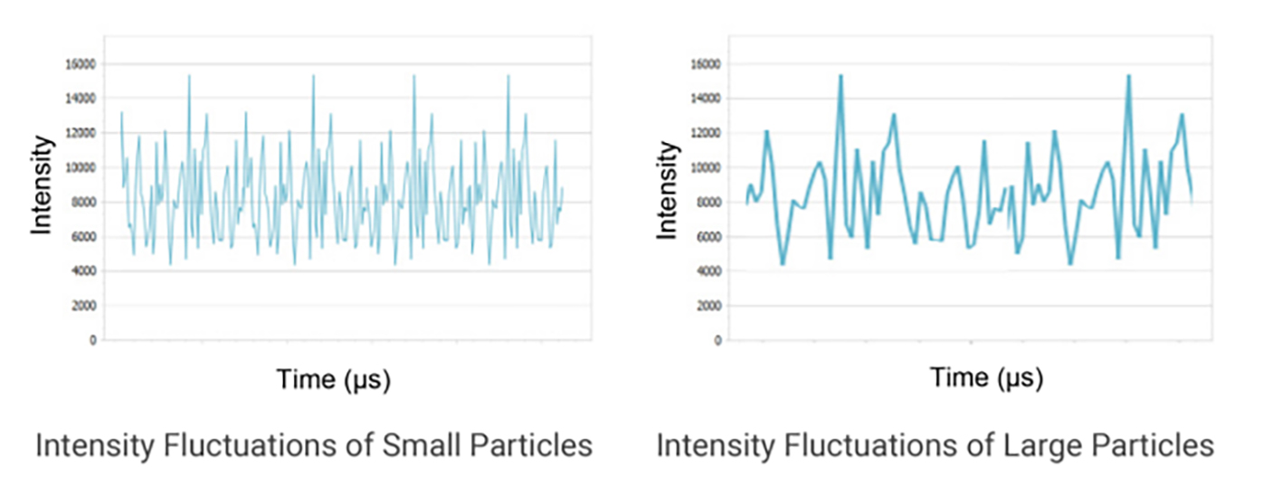
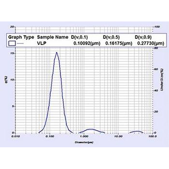
    Dynamic light scattering (DLS), also referred to as photon correlation spectroscopy (PCS) or quasi-elastic light scattering (QELS), is a technique used to measure Brownian motion in a dispersant. It is based on the principle that smaller particles move faster while larger particles move slower. The scattering intensities of the particles are detected by an avalanche photodiode (APD) and then converted into a correlation function using a correlator. From this correlation function, a mathematic algorithm can be applied to obtain the diffusion coefficient (D).


    3) Backscattering Detection Technology

    Features
    • Wider Concentration Range

      By optimizing the detection position, the highly concentrated samples can be detected near the edge of the sample cell, effectively minimizing the multiple light scattering effect.

    • Higher Sensitivity

      8-10 times scattering volume and around 10 times sensitivity as compared to the traditional 90° optical design.

    • Higher Size Upper Limit

      It mitigates multiple light scattering from large particles and, to some extent, reduces the number fluctuation of large particles due to the much larger scattering volume.
    • Better Reproducibility

      The DLS backscattering technology is less influenced by dust contaminants and unevenly distributed agglomerates and provides better reproducibility.
    Intelligent Search for the Optimal Detection Position

    The software automatically determines the optimal detection position based on the size, concentration, and scattering ability of the sample to achieve the highest measurement accuracy and offer flexibility in detecting different types and concentrations of samples. This feature is particularly useful when dealing with a variety of samples, each with its unique scattering properties and concentrations.

    4) Zeta Potential Measured by Electrophoretic Light Scattering (ELS)

    In aqueous systems, charged particles are surrounded by counterions that form an inner Stern layer and an outer shear layer. Zeta potential is the electrical potential at the interface of the shear layer. A higher zeta potential indicates greater stability and less aggregation of the suspension system. Electrophoretic light scattering (ELS) measures electrophoretic mobility via Doppler shifts of scattered light, which can be used to determine the zeta potential of a sample by Henry’s equation.

    5) Phase Analysis Light Scattering (PALS)

    Phase analysis light scattering (PALS) is an advanced technology based on the traditional ELS technology, which has been further developed by Bettersize to measure the zeta potential and its distribution of a sample.

    Features and Benefits
    • Accurate measurement of samples with low electrophoretic mobility
    • Effective for samples in organic solvents with low dielectric constant
    • More accurate results for samples with high conductivity
    • Effectively measures the zeta potential of particles whose charge approaches the isoelectric pointzeta potential distribution

    6) Static Light Scattering

    Static light scattering (SLS) is a technology that measures the scattering intensities, weight-average molecular weight (Mw), and second virial coefficient (A2) of the sample through the Rayleigh equation:

    where c is the sample concentration, θ is the detection angle, Rθ is the Rayleigh ratio used to characterize the intensity ratio between the scattered light and the incident light at angle θ, Mw is the sample’s weight-average molecular weight, A2 is the second virial coefficient, and K is a constant related to (dn/dc)2.

    During molecular weight measurements, scattering intensities of the sample at different concentrations are detected. By using the scattering intensity and Rayleigh ratio of a known standard (such as toluene), the Rayleigh ratios of samples at different concentrations are computed and plotted into a Debye plot. The molecular weight and the second virial coefficient are then obtained through the intercept and slope from the linear regression of the Debye plot.

    7) Microrheology Measured by DLS

    Dynamic Light Scattering Microrheology (DLS Microrheology) is an economical and efficient technique that utilizes dynamic light scattering to determine rheological properties. By analyzing the Brownian motion of colloidal tracer particles, information about the viscoelastic properties of the system, such as viscoelastic modulus, complex viscosity and creep compliance, can be obtained with the generalized Stokes-Einstein equation.

    Features & Benefits
    • Investigates rheological behaviors by measuring the thermally-driven motion of tracer particles within a material being studied
    • Facilitates the measurement across a wide range of frequencies
    • Applies low stress to tracer particles
    • Requires only a microliter-scale sample volume
    • Complements mechanical rheology results
    • Suitable for weakly-structured samples

    8) Temperature Trend Measurement

    Measurement Parameters
    • Size vs. Temperature
    • Zeta Potential vs. Temperature
    Features
    • Benefit protein formulation stability study
    • Accelerates real-time aging through elevated temperature simulation
    Benefits
    • Benefit protein formulation stability study
    • Accelerates real-time aging through elevated temperature simulation

    9) pH Trend Measurement

    Measurement Parameters
    • Zeta Potential vs. pH
    • Isoelectric point
    • Conductivity vs. pH
    Features
    • High-precision ternary titration pumps
    • Controllable peristaltic pump with high flow capacity and high flow rate
    • General-purpose electrode
    • Automated titrant selection based on initial and target pH using intelligent software
    Benefits
    • Completes measurements within a shorter time
    • Improves consistency and repeatability of results
    • Reduces the workload of researchers
    • Simplifies qualifications needed for operators
    • Accelerates real-time aging through elevated temperature simulation
    • Reduces exposure to corrosive liquids

    10) A Research Level Software

    • SOP guarantees measurement accuracy and completeness
    • Automatic calculation of mean and standard deviation for results and statistics
    • Comparison of results from multiple runs through statistics and overlay functions
    • Real-time display of information and results
    • Over 100 available parameters that meet research, QA, QC, and production needs
    • Free lifelong upgrades provided
    • 11) Compliance With FDA 21 CFR Part 11

      The BeNano software system is compliant with 21 CFR Part 11 regulations, which restricts access to authorized individuals through a username and password system for electronic record signing, access logs, change logs, or operation execution. An activation code can be used to upgrade security settings and ensure compliance, and an “audit trail” can be viewed to ensure proper management and maintenance of system security and data integrity.

  • Technology

  • Accessories

    BAT-1 Autotitrator

    1) Introduction

    The BAT-1 Autotitrator is equipped with three high-precision titration pumps (with precision of 0.28 μL), and a magnetic stirrer, and is in combination with the BeNano series nanoparticle size and zeta potential analyzer for automatic acid-base titration and determination of isoelectric point (IEP). The pinch valve can close the circuit of the sample during the measurement, leading to high efficiency, accurate titration, good repeatability and the results being independent of operators. The disposable sample container can avoid the sample cross-contamination.

    2) Features
    • Combination electrode with high precision and high feedback speed
    • High precision ternary titration pumps
    • Controllable peristaltic pump with high flow capacity and high flow rate
    • Internal magnetic stirrer system
    • SOP operation
    • Replaceable tubes
    • Corrosion resistant design
    • General purpose electrode
    • Intelligentization
    • Determination of isoelectric point
    3) How it works

    The BAT-1 Autotitrator is designed to be used with the BeNano series for the measurement of zeta potential over a wide pH range, providing the information of zeta potentials and the stability of samples in different conditions. The operation flow is as follows:

    • a). Preparing the samples to be detected and the titrants in the containers, respectively;
    • b). Creating or editing a titration SOP in BeNano software by setting the volume of the sample to be measured, the concentrations of the titrants, the initial pH, the target pH, the pH interval and the target pH tolerance, etc.;
    • c). To start the determination, the sample is titrated to approach to the first pH value through automatic calculation, and is injected into the folded capillary cell by the peristaltic pump for zeta potential measurement;
    • d). Repeating the above procedures until approaching the final target pH automatically;
    • Saving and outputting complete data and the trend plot of zeta potential vs. pH;
    • Giving the isoelectric point if it is included in the setting pH range.

    4) Download BAT-1 Autotitrator Flyer
  • Applications

    Battery Electrode Slurry

    Monosaccharide Molecules

    Resins

    BSA Solution

    Aluminium Oxide

    Titanium Oxide

    Thermal Sensitive Hydrogel

    Latex antibody immunological reagents

    Alumina Abrasives

    Iron Dextran

    Fat Emulsion

    BSA Solution

    Surfactant Micelles

    Pigment

    Bovine Serum Albumin

    Coffee Creamer Powder

    Lysozyme

  • Specification

    Functions

    Parameter

    BeNano 180 Zeta Pro

    BeNano 180 Zeta

    BeNano 90 Zeta

    BeNano Zeta

    BeNano 180 Pro

    BeNano 180

    BeNano 90

    Size
    measurement

    Size
    measurement range

    0.3 nm – 15 μm*

    0.3 nm – 10 μm*

    0.3 nm – 15 μm*

    N/A

    0.3 nm – 15 μm*

    0.3 nm -10 μm*

    0.3 nm – 15 μm*

    Sample volume

    3 μL – 1 mL*

    40 μL – 1 mL*

    3 μL – 1 mL*

    N/A

    3 μL – 1 mL*

    40 μL – 1 mL*

    3 μL – 1 mL*

    Detection angle

    90° & 173° & 12°

    173° & 12°

    90° & 12°

    N/A

    90° & 173°

    173°

    90°

    Analysis algorithm

    Cumulants, General Mode,
    CONTIN

    Cumulants, General Mode,
    CONTIN

    Cumulants, General Mode,
    CONTIN

    N/A

    Cumulants, General Mode,
    CONTIN

    Cumulants, General Mode,
    CONTIN

    Cumulants, General Mode,
    CONTIN

    Upper limit of
    concentration range

    40% w/v*

    40% w/v*

    Optically clear+

    N/A

    40% w/v*

    40% w/v*

    Optically clear†

    Detection position

    Movable position
    0.4 – 5 mm

    Movable position
    0.4 – 5 mm

    Fixed position
    5 mm

    N/A

    Movable position
    0.4 – 5 mm

    Movable position
    0.4 – 5 mm

    Fixed position
    5 mm

    Zeta potential
    measurement

    Detection angle

    12°

    12°

    12°

    12°

    N/A

    N/A

    N/A

    Zeta potential
    measurement range

    No actual limitation

    No actual limitation

    No actual limitation

    No actual limitation

    N/A

    N/A

    N/A

    Electrophoretic mobility

    > ± 20 μm·cm/V·s

    > ± 20 μm·cm/V·s

    > ± 20 μm·cm/V·s

    > ± 20 μm·cm/V·s

    N/A

    N/A

    N/A

    Conductivity

    0 – 260 mS/cm

    0 – 260 mS/cm

    0 – 260 mS/cm

    0 – 260 mS/cm

    N/A

    N/A

    N/A

    Sample volume

    0.75 – 1 mL

    0.75 – 1 mL

    0.75 – 1 mL

    0.75 – 1 mL

    N/A

    N/A

    N/A

    Sample size

    2 nm – 110 μm

    2 nm – 110 μm

    2 nm – 110 μm

    2 nm – 110 μm

    N/A

    N/A

    N/A

    Other
    measurements

    Molecular weight
    (Mw)

    342 Da – 2 x 107 Da* 342 Da – 2 x 10⁷ Da*

    342 Da – 2 x 10⁷ Da*

    N/A

    342 Da – 2 x 10⁷ Da*

    342 Da – 2 x 10⁷ Da*

    342 Da – 2 x 10⁷ Da*

    Viscosity

    0.01 cp – 100 cp*

    0.01 cp – 100 cp*

    0.01 cp – 100 cp*

    N/A

    0.01 cp – 100 cp*

    0.01 cp – 100 cp*

    0.01 cp – 100 cp*

    Interaction parameter
    KD

    No actual limitation

    No actual limitation

    No actual limitation

    N/A

    No actual limitation

    No actual limitation

    No actual limitation

    Trend measurement

    Time and temperature

    Time and temperature

    Time and temperature

    Time and temperature

    Time and temperature

    Time and temperature

    Time and temperature

    System
    parameters

    Temperature
    control range

    -15℃ – 110℃,
    ±0.1℃

    -15℃ – 110℃,
    ±0.1℃

    -15℃ – 110℃,
    ±0.1℃

    -15℃ – 110℃,
    ±0.1℃

    -15℃ – 110℃,
    ±0.1℃

    -15℃ – 110℃,
    ±0.1℃

    -15℃ – 110℃,
    ±0.1℃

    Condensation control

    Dry air or nitrogen

    Dry air or nitrogen

    Dry air or nitrogen

    Dry air or nitrogen

    Dry air or nitrogen

    Dry air or nitrogen

    Dry air or nitrogen

    Laser source

    50 mW Solid-state laser, 671 nm#, Class 1

    50 mW Solid-state laser, 671 nm#, Class 1

    50 mW Solid-state laser, 671 nm#, Class 1

    50 mW Solid-state laser, 671 nm#, Class 1

    50 mW Solid-state laser, 671 nm#, Class 1

    50 mW Solid-state laser, 671 nm#, Class 1

    50 mW Solid-state laser, 671 nm#, Class 1

    Correlator

    Up to 4000 channels,

    1011 linear
    dynamic range

    Up to 4000 channels,

    1011 linear
    dynamic range

    Up to 4000 channels,

    1011 linear
    dynamic range

    Up to 4000 channels,

    1011 linear
    dynamic range

    Up to 4000 channels,

    1011 linear
    dynamic range

    Up to 4000 channels,

    1011 linear
    dynamic range

    Up to 4000 channels,

    1011 linear
    dynamic range

    Detector

    Avalanche photodiode
    (APD)

    Avalanche photodiode
    (APD)

    Avalanche photodiode
    (APD)

    Avalanche photodiode
    (APD)

    Avalanche photodiode
    (APD)

    Avalanche photodiode
    (APD)

    Avalanche photodiode
    (APD)

    Intensity control

    0.0001% – 100%,
    manual or automatic

    0.0001% – 100%,
    manual or automatic

    0.0001% – 100%,
    manual or automatic

    0.0001% – 100%,
    manual or automatic

    0.0001% – 100%,
    manual or automatic

    0.0001% – 100%,
    manual or automatic

    0.0001% – 100%,
    manual or automatic

    Dimensions
    (L x W x H)

    62.5 x 40 x 24.5 cm
    (22 kg)

    62.5 x 40 x 24.5 cm
    (22 kg)

    62.5 x 40 x 24.5 cm
    (22 kg)

    62.5 x 40 x 24.5 cm
    (22 kg)

    62.5 x 40 x 24.5 cm
    (22 kg)

    62.5 x 40 x 24.5 cm
    (22 kg)

    62.5 x 40 x 24.5 cm
    (22 kg)

    Power supply

    AC 100-240 V,
    50-60 Hz, 4A

    AC 100-240 V,
    50-60 Hz, 4A

    AC 100-240 V,
    50-60 Hz, 4A

    AC 100-240 V,
    50-60 Hz, 4A

    AC 100-240 V,
    50-60 Hz, 4A

    AC 100-240 V,
    50-60 Hz, 4A

    AC 100-240 V,
    50-60 Hz, 4A

    Conformity
    to standards

    21 CFR Part 11, ISO 13321, ISO 22412, ISO 13099

    21 CFR Part 11, ISO 13321, ISO 22412, ISO 13099

    21 CFR Part 11, ISO 13321, ISO 22412, ISO 13099

    21 CFR Part 11, ISO 13321, ISO 22412, ISO 13099

    21 CFR Part 11, ISO 13321, ISO 22412, ISO 13099

    21 CFR Part 11, ISO 13321, ISO 22412, ISO 13099

    21 CFR Part 11, ISO 13321, ISO 22412, ISO 13099

    Optional Accessories

    Disposable
    micro-volume cuvette

    40 – 50 μL

    40 – 50 μL

    40 – 50 μL

    N/A

    40 – 50 μL

    40 – 50 μL

    40 – 50 μL

    Micro-volume
    glass cuvette

    25 μL

    N/A

    25 μL

    N/A

    25 μL

    N/A

    25 μL

    Glass cuvette
    with round opening

    1 mL

    1 mL

    1 mL

    N/A

    1 mL

    1 mL

    1 mL

    Capillary sizing cell

    3 – 5 μL

    N/A

    3 – 5 μL

    N/A

    3 – 5 μL

    N/A

    3 – 5 μL

    Dip cell kit

    1 – 1.5 mL,
    zeta potential measurement
    for organic-based samples

    1 – 1.5 mL,
    zeta potential measurement
    for organic-based samples

    1 – 1.5 mL,
    zeta potential measurement
    for organic-based samples

    1 – 1.5 mL,
    zeta potential measurement
    for organic-based samples

    N/A

    N/A

    N/A

    * Dependent on samples and accessories

    † Up to 40% w/v using capillary sizing cell

    # 10mW 633nm He-Ne laser available on request

Not sure if it’s the right instrument?

No worries, send us a sample and we will test it for you


Send a sample

Continue reading

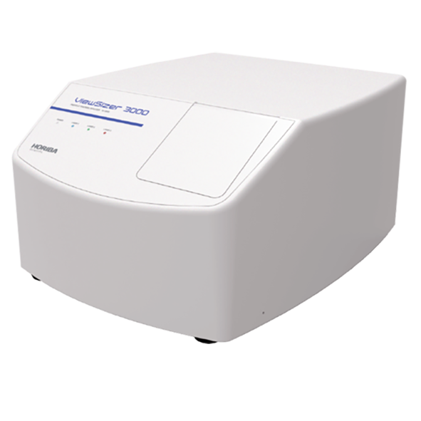
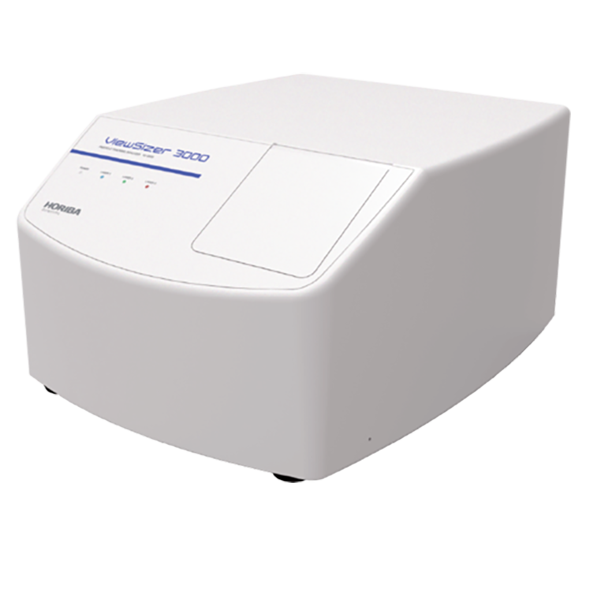
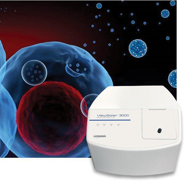
ViewSizer 3000

Horiba Scientific
ViewSizer 3000

Simultaneous Multi-Laser Nanoparticle Tracking Analysis (NTA)

  • Measurement range 10 nm – 15 µm
  • Concentration Measurement Range: 1E5 to 1E9 particles/mL
  • No cross-contamination

Download
brochure
   Request
quote

Beckman Coulter LS 13 320 XR Laser Diffraction Particle Size Analyser 21 CFR Part 11

Exosomes? Virus? Nanoparticle? Use multiple lasers for complete, detailed analysis of all the particles in your sample.

Exosomes, viruses, and nanoparticles all have wide size distributions which defeat traditional Nanoparticle Tracking Analysis (NTA) analyzers. The ViewSizer 3000 features simultaneous measurement with three lasers to collect the most accurate distribution and concentration information over a wide range of sizes within the same sample. Where the signal from a particle is too bright and saturates the detector from one laser, the software automatically uses data from a lower power laser to ensure the most accurate size and concentration information. On the other hand, when scattering from one laser is too weak for detection, the software uses data from a higher power laser to accurately track the particle.

Cross contamination is a concern in all analyses. Simplified cleaning means thorough cleaning. The easy-to-remove sample cells can be dissassembled for rapid, thorough cleaning, which leads to better data.

Escape the limits of traditional Nanoparticle Tracking Analysis

Accurate and sensitive analysis without cross contamination

Not sure if it’s the right instrument?

No worries, send us a sample and we will test it for you


Send a sample

Continue reading

VideoDrop

Myriade
VideoDrop

Nanoparticle Size and Concentration Analyser

  • Results in real time
  • Easy to use
  • No labeling
  • No purification

Download
brochure
   Request
quote

Beckman Coulter LS 13 320 XR Laser Diffraction Particle Size Analyser 21 CFR Part 11

  • Overview

    An ultra fast measurement
    thanks to 4 decisive advantages

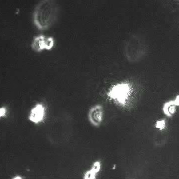
    Videodrop films in real time biological nanoparticles between 80 nm & 500 nm and provides a microscopic view up to 10µm in a drop. The compact device, assisted by a dedicated software, allows the manipulation to be very easy to perform. No labeling is necessary on the analyzed sample. It is possible to work on unpurified solutions and on little volumes (5µL) with a concentration range of 10^8 to 10^10 particles/ml.

    DEMONSTRATION

    Measure in a single drop the concentration and the size of nanoparticles
    in less than one minute ? Let’s go…

    The Ultimate Nanoparticle Analysis Solution: VideoDrop

    For precise nanoparticle size and concentration analysis, the VideoDrop excels with advanced video-based analysis, dynamic light scattering, and particle tracking. This ensures accurate and reliable measurements for viruses, extracellular vesicles, phages, lipids, and other nanomaterials.

    Key Benefits:

    • Precision & Accuracy: Delivers reliable data for confident research and applications.
    • High-Resolution Imaging: Offers detailed insights into nanoparticle morphology and behaviour.
    • Real-Time Analysis: Monitors particle dynamics in real-time, crucial for virology and other fields.
    • Versatility: Suitable for diverse applications across pharmaceuticals, biotechnology, and material science.
    • User-Friendly Interface: Simplifies operation, increasing productivity for all users.
    • Cost-Effective: Provides a robust, low-maintenance solution with high return on investment.
    • Advanced Software: Facilitates comprehensive data analysis and visualisation.
    • High-Throughput: Efficiently processes multiple samples for large-scale research and quality control.

    Choose VideoDrop for unparalleled nanoparticle analysis, enhancing your research and applications with precision and efficiency.

  • Technology

    Based on the principles of interferometry, the Videodrop allows you to « see »
    biological nanoparticles in the range of 80-500 nm.
    The observation is done in 3 steps.


  • Applications

    Viruses

    Extracellular Vesicles

    Phages and Lipids

    Nanoparticles

Not sure if it’s the right instrument?

No worries, send us a sample and we will test it for you


Send a sample

Continue reading

Moxi V

Orflo
Moxi V

Automated Cell Counter

  • True Cell Viability Counts
  • Precision Sizing
  • Highly Accurate
  • Rapid Assays

Download
brochure
   Request
quote

Beckman Coulter LS 13 320 XR Laser Diffraction Particle Size Analyser 21 CFR Part 11

Gold Standard Automated Cell Counter, Cell Size, and Viability

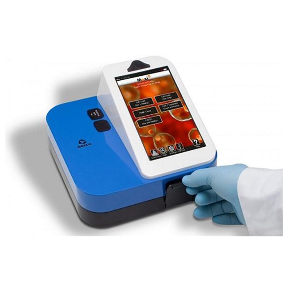
The Orflo Moxi V is your top choice for an Automated Cell Counter due to its exceptional accuracy and efficiency. Utilising Coulter Principle and fluorescence-based technology, it provides precise, reliable results quickly. Its user-friendly interface and high-throughput capability streamline operations, making it ideal for research. With its compact design and robust performance, the Moxi V ensures consistent, high-quality cell analysis, making it a valuable addition to any laboratory.

  • Overview

    The Moxi V Automated Cell Counter provides a combination of volumetric cell sizing (Particle Sizer) with simultaneous fluorescence (Cell Analyzer) to provide the most accurate cell counts, size, and viability in the industry. Specifically, the Moxi V employs the Coulter Principle to precisely measure the volumetric particle size of each particle for exact size measurements down to 3um in diameter (14fL volume), easily distinguishing between cells and debris. The system is also equipped with a 532nm laser and a 561nm/LP detection channel for robust cell viability analysis. Propidium Iodide (PI)-stained dead cells measure 50-100 times brighter fluorescence on the system than do live cells, removing the ambiguity associated with traditional Trypan Blue viability assessments. For each test, these size and viability measurements are applied, individually, to up to 23,000 cells in a matter of a few seconds. This ensures the highest level of precision and statistical robustness. As the fluidic volume is precisely metered as well, the particle counts are presented as an exact cell concentration.

    The Moxi V employs a patented, single-use, microfluidic flow cell. The flow cells eliminate the hassle of traditional Cell Analyzers and Coulter Counters, eliminating the need for cleaning, maintenance, clearing of clogs, cross contamination and occasionally replacement of bottles and tubes. The Moxi V uses very little sample volume, 60ul, allowing you to conserve your precious, potentially expensive, sample (e.g. stem cells). Cell concentrations as low as 10 cells/ul are possible, typically requiring just 5ul of cell sample diluted in 55ul of PBS.

    The Moxi V™ system comes standard with an ultra-intuitive, plug-and-play interface with free OS updates for as long as you own the instrument. No prior Cell Analysis experience is required – you simply just plug and play.

    Some Key Features of the Moxi V include:
    • True Cell Viability Counts – 50-100x more sensitive than vision counters.
    • Precision Sizing – Uses the Coulter Principle to get precise cell volumes with CVs less than 3% and no need for triplicates.
    • Highly Accurate – Accurate counts for smaller cells down to 3um (i.e. nuclei, RBCs). Uniquely accurate at low cell concentrations down to 10 cells per ul.
    • Rapid Assays – Offers a less than 15 second test that counts up to 23,000 cells compared to 200-300 counts on most vision counters.
  • Technical Specs

    Id

    MXV102

    Included accessories

    USB power cord, US style USB power adapter, and Type S+ cassette pack

    AC power type

    110 VAC

    Applications

    Gold Standard Cell Count, Cell Size, and Viability

    Battery Type

    Rechargeable 3.7 V, 7500 mAh lithium ion

    Cassette types

    Type S+

    Cell Particle Concentration Range

    10,000 – 5,000 cells/mL Type S+

    Cell types tested

    HEK-293
    HeLa
    PC12
    CD3+T
    CHO-K1
    Cos-7
    HepG2
    Hybridoma
    Jurkat E6-1

    MCF7
    Mesenchymal SC
    Monocyte
    Mouse ESC
    NIH 3T3
    PBMC (cultured)
    Red Blood Cells (RBC)
    L5178y
    C. albicans (Yeast)
    S. cerevisiae Vin 13 (Yeast)
    S. cerevisiae X5 (Yeast)
    Wine Yeast (natural fermentaion)
    S.cerevisiae (Baker’s Yeast
    Safale US-05 Yeast

    Data output formats

    FCS 3.1, screen shots (.bmp), CSV

    Data storage capasity

    4Gb

    Display resolution

    800 x 480 colour touchscreen

    Excitation wavelengths

    532nm

    In British units

    8 lbs

    Intended use statement

    For Research Use Only. Product is not for use in diagnostic procedures

    Laser colour

    Green

    Measurable dynamic range

    3 – 27 microns Type Type S+
    4 – 35 microns Type MF-M

    Measurement time

    10 seconds Type S+

    MPI cell health ratio

    Yes (Size histogram only)

    Number of detection channels flow parameters

    2 colour, 1 size, 1 forward extinction

    Number of PMTs

    1

    Optical detection range

    561nm/LP (e.g. PI)

    Particle size detection method

    Impedimetric (Coulter Principle)

    Pre-programmed tests

    Gold Standard Cell Count, Cell Size, and Viability

    Sample type

    Cell Preparations

    Sample volume

    60 µL

    Supported connectivity

    USB on-the-go

    Useable cell volume

    14 – 10,306 fL Type S+
    14 – 22,449 fL Type MF-M

    Weight

    3.6 kg

  • How it works

    The operating principle behind the Moxi V is a unique combination of Coulter-Principle cell size determination with simultaneous fluorescence detection. As cells flow single file through the microfabricated single-use flow cell the volume of each particle is measured at the exact same time as their primary fluorescence is measured using a 532nm solid state diode laser with a 561nm/LP (e.g. PI) emission filter. Thousands of cells are measured in the 10 second read time and the data are plotted in a gradient density scatter plot as Cell size (volume) vs. Fluorescence (PMT voltage). Gating is automatically performed by the system but can be easily adjusted/tuned by the user. The resulting live/dead ratios are automatically calculated (depending on the app selected). The analyzed data can also be displayed as a two color size histogram. Total volumetric cell counts are automatically determined for each test by precisely measuring the volume of fluid being analysed.

    Step 1:
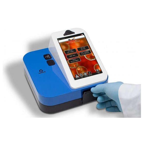
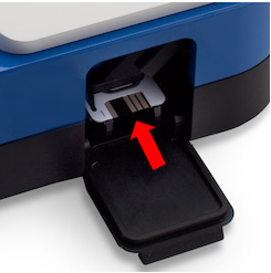
    Select desired app, insert the cassette and close the doors.

    Step 2:
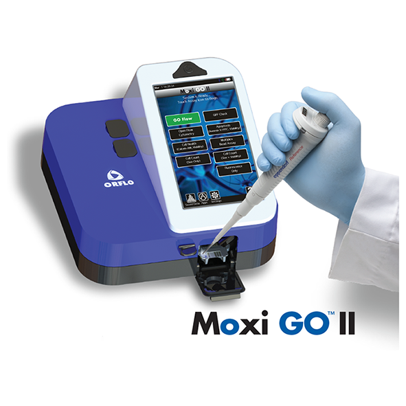
    Once auto-alignment is complete, open the top door and pipette 60μl of labelled sample into the cassette.
    Step 3:
    Close the top door, assays run automatically and results are generated in ~10 seconds. Note: Each cassette holds 2 tests. When Sample 1 is complete, simply re-insert other end of cassette into Moxi V, and load Sample 2.
    Data

    Data can be displayed on the unit in both a color density scatter plot and a two color size histogram. Simply drag gates using the intuitive touch display for instant live/dead ratio calculations and each of the gated volumetric cell counts (i.e., total population, live population, and dead population (Viability App). The mean cell volume for the gated populations is also automatically displayed on the unit. Results from each test are stored in the standard FCS 3:1 format and can be viewed using any Cell Analysis package. The actual Moxi V screenshots from each assay (dot plots and histograms) are also stored in bitmap format for use online. Hundreds of files can be stored on each Moxi V and are easily transferred to a Mac or PC using USB on-the-go. No aditional software is required.

Not sure if it’s the right instrument?

No worries, send us a sample and we will test it for you


Send a sample

Continue reading

Moxi GO II

Orflo
Moxi GO II

Mini Automated Cell Analyzer

  • Small footprint enables portability and convenience.
  • Fast (<10 sec) test times and no warm-up periods ensure rapid results.
  • Intuitive, touchscreen design for ease of use.
  • No cleaning or maintenance required.

Download
brochure
   Request
quote

Beckman Coulter LS 13 320 XR Laser Diffraction Particle Size Analyser 21 CFR Part 11

Next Generation Coulter-Principle Cell Analyser.

The Orflo Moxi GO II  Cell Analyser is the best solution for cell counting due to its precision, speed, and ease of use. Combining Coulter Principle-based sizing with fluorescence-based viability, it offers highly accurate and reproducible results. Its intuitive interface and automated processes ensure rapid and user-friendly operation, making it ideal for both novice and experienced users. The Moxi GO II’s compact design and high-throughput capabilities make it perfect for various applications, including research labs and biotech industries, providing consistent and reliable cell analysis.

  • Overview

    Combines the Coulter Principle (for highly-accurate cell counts and exact, volumetric cell sizing) with a 488nm laser and two PMT fluorescence detection channels (for cell health assays, robust CAR-T monitoring, cellular response profiling, and immuno-profiling).

    The Moxi GO II’s new Auto-Gating feature will analyse results in an accurate, repeatable way to provide the most consistent results — presented in a simplified new Data Summary page. The new Batch Mode feature allows you to run multiple tests of the same sample type. It will auto-find live and dead cell populations, eliminating user-to-user variability.

  • Technical Specs

    Id

    MXG102

    Included accessories

    USB power cord, US style USB power adapter, and Type S+ cassette pack

    AC power type

    110 VAC

    Applications

    Mulitplexed Bead ELISA’s
    In Cell Westerrns
    In Cell Protein Quant
    GFP
    Gold Standard Cell Count and Viability
    Mito Potential
    ROS
    Phagocytosis

    Battery Type

    Rechargeable 3.7 V, 4400 mAh lithium ion

    Cassette types

    Type S+

    Cell Particle Concentration Range

    5,000 – 1,000,000 cells/mL Type S+

    Cell types tested

    HEK-293
    HeLa
    PC12
    CD3+T
    CHO-K1
    Cos-7
    HepG2
    Hybridoma
    Jurkat E6-1
    K562
    MCF7
    Mesenchymal SC
    Monocyte
    Mouse ESC
    NIH 3T3
    PBMC (cultured)
    Red Blood Cells (RBC)
    L5178y
    C. albicans (Yeast)
    S. cerevisiae Vin 13 (Yeast)
    S. cerevisiae X5 (Yeast)
    Wine Yeast (natural fermentaion)
    S.cerevisiae (Baker’s Yeast
    Safale US-05 Yeast

    Data output formats

    FCS 3.1, screen shots (.bmp), CSV

    Data storage capasity

    4Gb

    Display resolution

    800 x 480 color touchscreen

    Excitation wavelengths

    488nm

    In British units

    10 lbs

    Intended use statement

    For Research Use Only. Product is not for use in diagnostic procedures

    Laser colour

    Blue

    Measurable dynamic range

    3 – 27 microns Type Type S+
    4 – 35 microns Type MF-M

    Measurement time

    10 seconds Type S+

    MPI cell health ratio

    Yes (Size histogram only)

    Number of detection channels flow parameters

    2 color, 1 size, 1 forward extinction

    Number of PMTs

    2

    Optical detection range

    525/45nm (e.g. FITC, GFP) and 561nm/LP (e.g. PE, RFP)

    Particle size detection method

    Impedimetric (Coulter Principle)

    Platform

    Open platform: 561nm/LP (PI, PE, DS Red, Sytox Orange, 7 AAD, Nile Red, Rhodamine Red, Sun Coast Yellow, PE/Cy5), 525/45nm (FITC, GFP, Alexa Fluor 488nm, Calcein)

    Pre-programmed tests

    Mulitplexed Bead ELISA’s
    In Cell Westerrns
    In Cell Protein Quant
    RFP
    Gold Standard Cell Count and Viability
    Mito Potential
    ROS
    Phagocytosis

    Resolution histogram bins

    1000

    Sample type

    Beads
    Cell Preparations

    Sample volume

    60 µL

    Supported connectivity

    USB on-the-go

    Useable cell volume

    14 – 10,306 fL Type S+
    14 – 22,449 fL Type MF-M

    Weight

    4.53 kg

  • How it works

    The operating principle behind the Moxi GO II Cell Analysers is a unique combination of Coulter-style cell size determination with simultaneous fluorescence detection. As cells flow single file through the microfabricated single-use flow cell the volume of each particle is measured at the exact same time as their primary fluorescence is measured using a 488nm (MXG102) solid state diode laser with and with the following emission filters – 525/45nm (e.g. FITC, GFP, Alexa 488) and 561nm/LP (e.g. PE, RFP). Thousands of cells are measured in the 10 second read time and the data are plotted in a gradient density scatter plot as Cell size (volume) vs. Fluorescence (PMT voltage). Gating is easily performed on the unit using a interactive touch display, and the resulting live/dead ratios are automatically calculated (depending on the app selected). The analyzed data can also be displayed as a two color size histogram. Total volumetric cell counts are automatically determined for each test by precisely measuring the volume of fluid being analysed.

    Step 1:
    Select desired app, insert the cassette and close the doors.

    Step 2:
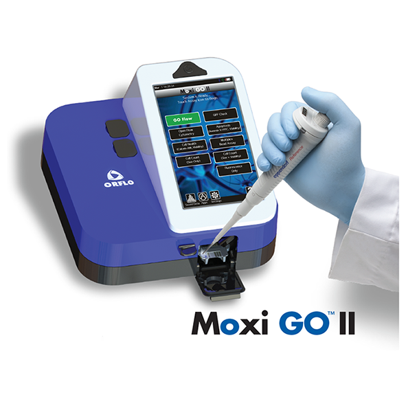
    Once auto-alignment is complete, open the top door and pipette 60μl of labelled sample into the cassette.
    Step 3:
    Close the top door, assays run automatically and results are generated in ~10 seconds. Note: Each cassette holds 2 tests. When Sample 1 is complete, simply re-insert other end of cassette into Moxi GO, and load Sample 2.
    Data

    Data can be displayed on the unit in both a color density scatter plot and a two color size histogram. Simply drag gates using the intuitive touch display for instant live/dead ratio calculations and each of the gated volumetric cell counts (i.e., total population, live population, and dead population (Viability App). The mean cell volume for the gated populations is also automatically displayed on the unit. Results from each test are stored in the standard FCS 3:1 format and can be viewed using any Cell Analysis analysis package. The actual Moxi Flow screenshots from each assay (dot plots and histograms) are also stored in bitmap format for use online. Hundreds of files can be stored on each Moxi GO and are easily transferred to a Mac or PC using USB on-the-go. No aditional software is required.


  • Applications

    Apoptosis Monitoring

    Transfection Monitoring

    Yeast Monitoring in Brewing

    Immuno-phenotyping

    Mitochondrial Membrane

    Reactive Oxygen Species

Not sure if it’s the right instrument?

No worries, send us a sample and we will test it for you


Send a sample

Continue reading

Moxi Z

Orflo
Moxi Z

Mini Automated Cell Counter Kit

  • Produce repeatable cell counts with >95% accuracy in just 8 seconds
  • Improve downstream results with better quality control
  • Offers Precise Cell Counting
  • Assess cell culture health without reagents or dyes

  • Measure the smallest cells at very high concentrations

Download
brochure
   Request
quote

Beckman Coulter LS 13 320 XR Laser Diffraction Particle Size Analyser 21 CFR Part 11

MOXI Z Mini Automated Cell Counter Kit – Coulter Principle Counts and Sizing. 

The Orflo Moxi Z offers precise cell counting through its advanced Coulter Principle technology and fluorescence-based viability analysis. Its user-friendly interface ensures quick and accurate measurements, making it ideal for research and clinical settings, enhancing cell analysis efficiency with reliable, reproducible results for various applications.

For research Use Only. Not for use in diagnostic procedures.

  • Overview

    Improve downstream results with better quality control.

    Moxi Z is the only automated cell counter that combines the Coulter Principle typically used in high-end cell counters with a patented thin-film sensor technology to allow for highly accurate (> 95%) and repeatable particle counting and sizing for a broad range of cell types – from mammalian cells to cells as small as wine yeast and more. Since today’s workflows demand accurate quality control of samples, determining cell counts precisely has a significant impact on outcomes and downstream costs.

    Produce repeatable, precise cell counting with >95% accuracy in just 8 seconds.

    This ultra-small instrument uses patented microfluidic thin-film cassettes that enable automatic load and measure operation. The resulting single cell volumetric measurements are completely technique-independent. It provides the ideal alternative to the tedious manual counting associated with cytometers, or the inaccurate results associated with image-based automated cell counters (typical accuracies of 75-80%).

    Assess cell culture health without reagents or dyes.

    Moxi Z also automatically reports a unique cell health assessment index – MPI (Moxi Population Index) – without the need for reagents or dyes.MPI is a ratio of the cell population of interest relative to the entire particle distribution in that sample, factoring in dead cells that have shrunken or broken apart as well as other debris and contaminants in the sample. For monodisperse mammalian cultures this is closely correlated to the overall health of the sample.In the case of mixed cell populations, it’s provides an assessment of the relative fraction of the largest sized population in the sample. Unlike staining-based viability methods that focus on the uptake by dead cells and do not account for contaminants and other debris, MPI provides insight on the primary population of interest (based on size) and its relationship to ALL other particles in the sample.

    Measure the smallest cells at very high concentrations.

    Using the new Type S cassette, you can now accurately measure the smallest cells of any automated cell counter (down to 3um average diameter) at concentrations of up to 2.5e+6/ml. This means the new Type S cassette can measure Yeast (including wine yeast) and non-spherical particles down to 14 fL in volume.

    Results in just 8 seconds
    • Ultra-small and ultra-easy to use (no manual counting, no focusing of any kind)
    • Complete, high-resolution cell size/count histogram
    • Handles “de-aggregation” and coincidence events
    • Post-processing/analysis of count distributions with user-adjustable regions/gates
    • Assess cell health (MPI) without the need for reagents like Trypan blue
  • Technical Specs

    Id

    MXZ001

    Included accessories

    Instrument only

    AC power type

    110 VAC

    Applications

    Cell Counting | Cell Size | Cell Health

    Average Cell Diameter Range

    4 – 25 microns Type M | 3 – 20 microns Type S

    Battery Type

    Rechargeable 3.7 V, 4400 mAh lithium ion

    Cassette types

    Type M | Type S

    CE Certification

    Yes

    Cell Particle Concentration Range

    3,000 – 500,000 cells/mL Type M | 3,000 – 1,750,000 cells/mL Type S

    Cell types tested

    HEK-293
    HeLa
    PC12
    CD3+T
    CHO-K1
    Cos-7
    HepG2
    Hybridoma
    Jurkat E6-1
    K562
    MCF7
    Mesenchymal SC
    Monocyte
    Mouse ESC
    NIH 3T3
    PBMC (cultured)
    Red Blood Cells (RBC)
    L5178y
    C. albicans (Yeast)
    S. cerevisiae Vin 13 (Yeast)
    S. cerevisiae X5 (Yeast)
    Wine Yeast (natural fermentaion)
    S.cerevisiae (Baker’s Yeast
    Safale US-05 Yeast

    Data output formats

    .csv format (Orflo MoxiChart or Microsoft Excel)

    In British units

    1.5 lbs

    Intended use statement

    For Research Use Only. Product is not for use in diagnostic procedures

    Measurable dynamic range

    4- 34 microns Type M
    3 -26 microns Type S

    Measurement time

    15 seconds Type S
    10 seconds Type M

    MPI cell health

    Yes

    Number of detection channels flow parameters

    1

    Overall dimensions

    7.6 L x 4.3 W x 2.8 H (in)

    Particle size detection method

    Impedimetric (Coulter Principle)

    Platform

    No

    Pre-programmed tests

    None

    Sample type

    Mammalian Cells|Large Yeast|Large Algae|Protozoa Type M
    Mammalian Cells|Most Yeast|Small Algae|Small Protozoa Type S

    Sample volume

    75 µL

    Supported connectivity

    USB on-the-go; PC or Mac compatible; requires Windows XP, Windows Vista, Windows 7, or Mac OS X operating system

    Useable cell volume

    34 – 20,580 fL Type M
    14 – 4,200 fL Type S

    Weight

    0.68 kg

  • How it works

    Electronic current is passed through a small hole (Cell Sensing Zone) in a thin-film membrane. Cells flow substantially single file through the CSZ causing momentary increases in measured voltage that are directly proportional to cell or particle volume. Thousands of cells are measured during a single test and the size of each cell is plotted and saved in histogram format. Total volumetric counts are determined by precisely measuring the
    volume of fluid being analysed.

    Insert cassette into Moxi Z.

    Pipette 75 μL sample into cassette. Touch Screen.

    Finished! Automatic, hands-free measurement in just 8 seconds.

    Note: Each cassette holds 2 tests. When Sample 1 is complete, simply re-insert other end of cassette into MoxiZ, and load Sample 2.

    Data

    Data For each measurement, a complete histogram is displayed as well as cell count, average cell size and Moxi Population Index. The raw waveform from the most recent cell count, containing each cell spike, is also stored on the Moxi Z and can be both viewed on the Moxi Z and uploaded to the PC for additional analysis. The Moxi Z also performs sophisticated curve fitting analysis to more accurately determine cell counts in the event of overlapping populations of cells (or debris), significantly improving the accuracy of the volumetric counts produced by the Moxi Z. Data for 500 samples may be stored on the unit. All data can be transferred to a PC/Mac via Bluetooth. No special software is required for the PC since date files are Excel®-compatible. Sophisticated PC/Mac compatible analys software is included with each Moxi Z. Cell health may be assessed by comparing differences in histograms from culture to culture.

    Instructional Videos


  • Applications

    Blood Cells

    Monitoring Cell Health

    Algal Growth

    Yeast Monitoring

Not sure if it’s the right instrument?

No worries, send us a sample and we will test it for you


Send a sample

Continue reading

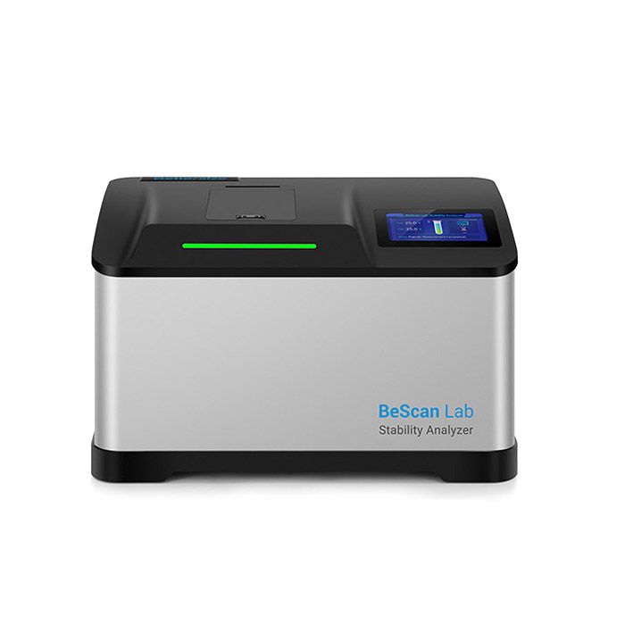
BeScan Lab

Bettersize BeScan

Stability Analyser

  • Particle  range 0.01 to 1,000 μm
  • Non-destructive stability analysis
  • Quantification of destabilisations and study of kinetics

Download
brochure
   Request
quote

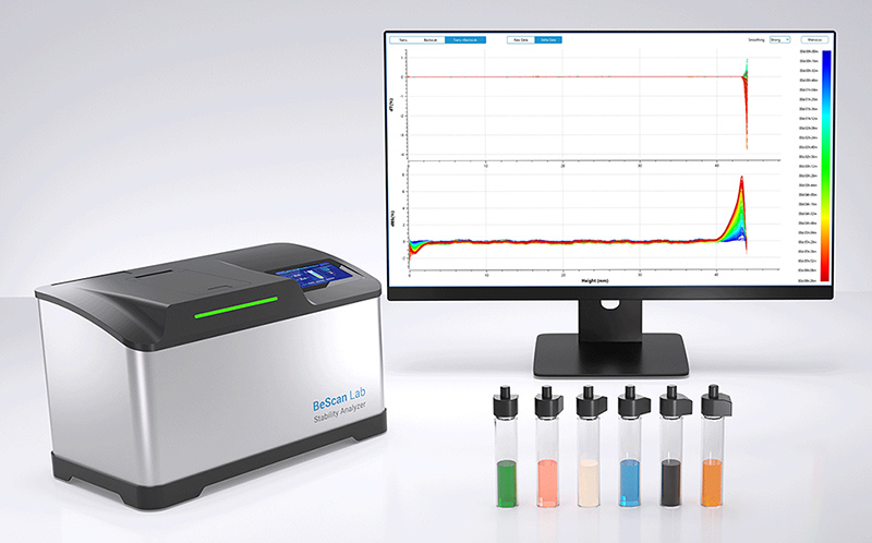
BeScan Lab Stability Analyser

BeScan Lab, the versatile, sensitive, and reliable stability analyser based on Static Multiple Light Scattering (SMLS) technology, is widely used in the formulation development and product quality control. It accommodates a wide range of sample concentrations up to 95% v/v and types such as emulsions, suspensions, and foams, with temperature scanning capabilities reaching up to 80 °C. BeScan Lab provides both qualitative analysis and quantification of destabilisation, helping you monitor long-term product stability and achieve optimal shelf life.

Features and Benefits

● Real stability analysis for dispersions with volume fraction up to 95%

● Particle size measurement range from 0.01 to 1,000 μm

● Non-destructive stability analysis: Non-contact, non-dilution, non-shearing

● 20 µm resolution data acquisition enables quicker sample stability observation than the naked eye

● Temperature control up to 80 °C to accelerate destabilisation

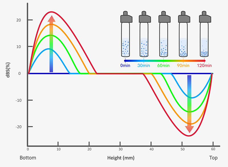
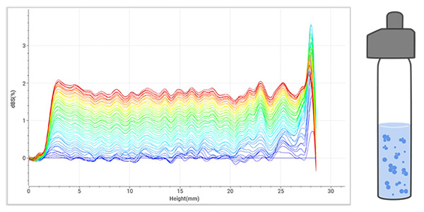
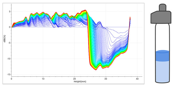
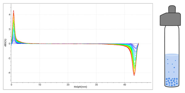
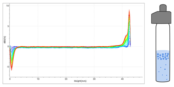
● Identification of various unstable phenomena: creaming, sedimentation, flocculation, coalescence, and phase separation

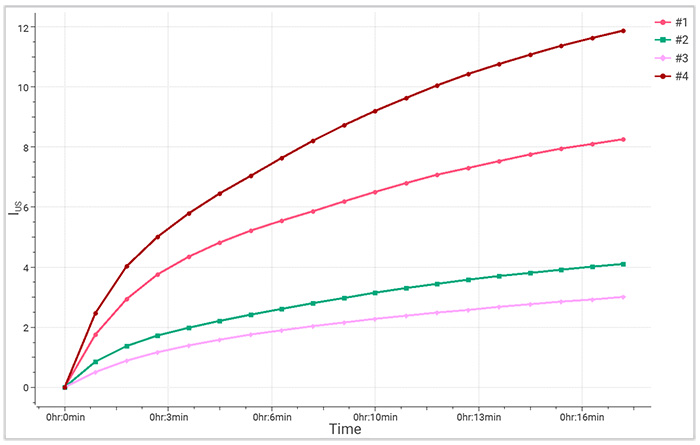
● Quantification of destabilisations and study of kinetics

  • Key Features

    What BeScan Lab Provides? 

    • lnstability index (lus)
    • Mean particle size
    • Hydrodynamic analysis
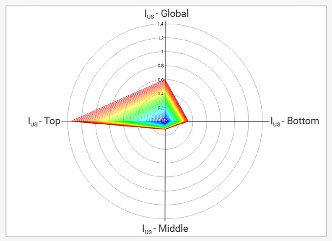
    • Radar chart for regional lus
    • Temperature trend testing
    • Particle migration rate

    Why You Need It? 

    From Raw Materials to Finish

    BeScan Lab plays a crucial role throughout the product lifecycle, supporting formulation, production, and pre-use stages. It enables formulation optimization, quality control during manufacturing, investigation into optimal transportation and storage conditions, and research on redispersibility

    1. Research and development

    Ensure excellent dispersibility and uniformity through raw material selection.

    2. Production and quality control

    Optimize production processes, including method, time, and temperature, to enhance efficiency.

    3. Storage and transportation

    Evaluate formulation stability under varying environmental conditions, observing destabilization, and predict shelf life.

    4. Pre-use treatment

    Study the reversibility of destabilization and compliance with usage standards.

    Features & Benefits

    Non-destructive stability analysis for various dispersions

    • Non-contact, non-dilution, non-shearing 
    • Sample volume fraction up to 95%
    • Particle size measurement range from 0.01 to 1,000 μm

    Fast and direct stability measurement

    • The high-performance LED and ultra-sensitive detectors, with a 20-micron scan step, allow real-time monitoring and capture of subtle variations 200 times faster than the naked eye
    • Temperature control up to 80 °C to accelerate destabilization

    Qualitative and quantitative stability results

    • Identification of various unstable phenomena, such as creaming, sedimentation, flocculation, coalescence, and phase separation
    • Quantification of destabilizations and study of kinetics


    Advanced Measurement Principle 

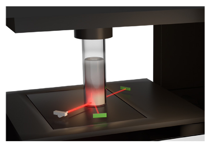
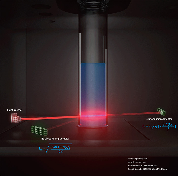
    Static Multiple Light Scattering (SMLS) is employed to characterize the stability of dispersions. Within BeScan Lab, a setup comprising two detectors and an LED light source ascends along the sample cell to conduct sample scanning. In the case of concentrated samples, the backward detector is employed to detect backscattered signals, while for diluted samples, the forward detector is utilized to detect transmitted signals.

    how-BeScan-Lab-woks

    Versatile Applications 

    • Agrochemicals

    Evaluate the stability of pesticide formulations to predict shelf life and ensure the consistent performance of suspension systems.

    • Battery and Energy

    Test the stability of electrode materials and electrolytes, crucial for enhancing battery performance and lifespan.

    • Ceramics

    Analyze the stability of ceramic slurries and monitor the stability of glazes and pigments, ensuring reliable production processes.

    • Home and Personal Care

    Ensure product stability in cosmetics, lotions, creams, and other formulations for reliable performance.

    • Food and Beverage

    Test the stability of food products, from milk to sauces, and assess the dispersibility of food powders to maintain product quality.

    • Petrochemicals

    Monitor and ensure the stability of oil products, providing critical insights into the long-term performance of lubricants and the behavior of polymers in oil.

    • Pharmaceuticals

    Conduct stability testing for medicinal formulations, assess long-term drug stability, and analyze biomacromolecule aggregation to ensure product efficacy.

    • Paints, Coatings and Inks

    Measure the stability of coatings and inks, and evaluate the dispersion of pigments and dyes for uniform product quality.

  • Technology

    Static Multiple Light Scattering  

    Static Multiple Light Scattering (SMLS) is an optical technique used to directly characterize native concentrated liquid dispersions. This technique emits light into the sample, where it is scattered multiple times by particles or droplets before being detected.

    BeScan Lab applies SMLS using an 850 nm LED as light source, with detectors set at 0° for capturing transmitted light and at 135° for backscattered light. This setup scans the sample vertically, analyzing the transmitted light for transparent systems, while the backscattered light is ideal for opaque systems.

    The signals are collected at 20 μm intervals, which enables precise observation of changes in size (d) and concentration (Φ) of suspended materials.

    Signal display

    Customized scanning procedures allow presentation of scans with different colors corresponding to different scanning times. The overlap of scans demonstrates how signals diverge from the reference as they vary with height and time. Intuitively, the scans capture local changes that characterize unstable phenomena.

    BeScan-Lab-Signal-display


    The example illustrates that during sedimentation, the backscattered signals (dBS) undergo a distinctive pattern of change: a decrease at the top and an increase at the bottom, which is attributed to the migration of particles.

    Features

    • Versatile measurement

    No limitations on color or viscosity, and suitable for a wide range of samples from low to high concentrations (up to 95% v/v).

    • Non-destructive and in situ 

    Measures without preparation, thus preserving the sample’s original characteristics.

    • Wide particle size range

    Capable of measuring particles from 0.01 to 1,000 μm.

    • Applicable to various systems

    Suitable for emulsions, suspensions, foams, and other dispersions, providing robust and high-resolution measurements.

    BeScan-Lab-Measurement-Principle

  • Software

    Dedicated Software 

    for Superior Qualitative and Quantitative Stability Outcomes

    Qualitative Analysis – Identification of Destabilisation

    BeScan Lab utilises near-infrared light and a precise 20-micrometer spatial resolution to detect early-stage destabilisation phenomena like phase separation, sedimentation, creaming and aggregation (flocculation, coalescence, and coagulation) well before they are visually observable.

    1. Flocculation often results in uniform changes in transmitted or backscattered signals across the entire sample height.

    • Common in wastewater treatment, electrode slurries, and drilling fluids.

    Data-results-of-Flocculation-analyzed-by-BeScan-Lab

    2. Phase separation typically involves evolving interfaces between phases over time.

    • Common in paints and coatings, cosmetics.

    Data-results-of-Phase-separation-analyzed-by-BeScan-Lab

    3. Sedimentation causes a decrease in backscattered signals at the top and an increase at the bottom in opaque samples.

    • Common in slurries, pigments, pesticides, vaccines, and body lotions.

    Data-results-of-Sedimentation-analyzed-by-BeScan-Lab

    4. Creaming in opaque samples enhances backscattered signals while lowering bottom signals.

    • Common in milk-based beverages, lipid emulsions, and pesticides.

    Data-results-of-Creaming-analyzed-by-BeScan-Lab

    Quantitative Analysis – Instability Index for Rating Guide

    BeScan Lab provides the instability index (IUS), which quantifies the stability of dispersions. The calculation involves summing all signal variations across the entire sample height and over time, capturing all subtle variations within the sample. This facilitates sample comparison, as a greater instability index (IUS) indicates lower stability. An instability index is automatically calculated after every scan using the following formula:

    instability-index-formula

    BeScan Lab offers instability indices over time to compare the stability of different samples. A slower increase in the instability index indicates higher dispersion stability, resulting in a flatter curve. Analysing the trend allows for predicting long-term stability.

    Time-dependent-instability-index

    Time-dependent instability index

    1. Phase separation dynamics and mean particle size

    Hydrodynamic analysis reveals layer thickness and particle migration rate over time, thereby determining the hydrodynamic mean diameter.

    Phase-separation-dynamics-and-mean-particle-size

    2. Optical analysis and mean particle size variation

    Particle size variation analysis is achievable with BeScan Lab, correlating transmitted and backscattered light signals.

    Optical analysis and mean particle size variation Particle size variation analysis is achievable with BeScan Lab, correlating transmitted and backscattered light signals.

    3. Temperature trend measurement

    Programmable temperature trend measurement up to 80°C, which explores stability under extreme conditions and accelerates destabilisation.

    Temperature-trend-measurement

    4. Radar chart

    Global and regional instability indices for each scanning are illustrated in form of a radar chart, intuitively providing a way to investigate regional stability (top, middle, and bottom).

    Radar-chart

  • Specification

    Parameters Values
    Measurement principle SMLS (Static Multiple Light Scattering)
    Detection angle 0° transmission and 135° backscattering
    Light source 850 nm LED
    Scan step 20 μm
    Scan height 0 – 60 mm
    Number of samples 1
    Maximum volume fraction* 95%
    Measurement range of particle size 0.01 – 1000 μm
    Temperature range RT – 80 ℃ (±0.5 ℃ )
    Sample volume 4 – 25 mL
    Measurement mode Regular/Fixed point/Temp. trend
    Dimension 460(L) x 260(W) x 280(H) mm
    Weight 13.5 kg
    Power AC100 – 240 V, 50 – 60 Hz, 3.8 A
    ISO compliance

    ISO/TR 18811:2018, ISO/TR 13097:2013

    ISO/TR 21357:2022, ISO/TS 22107:2021

     * Sample and sample preparation dependent

  • Applications

    Application

    Application

    Application

    Application

    Application

    Application

    Application

    Application

    Application

    Application

Not sure if it’s the right instrument?

No worries, send us a sample and we will test it for you


Send a sample

  • Name of customer

    Morbi ornare magna nec tortor rutrum interdum. Donec interdum facilisis elit, et iaculis nunc facilisis vitae. Sed quis semper dolor, id efficitur ex.
  • Name of customer

    Morbi ornare magna nec tortor rutrum interdum. Donec interdum facilisis elit, et iaculis nunc facilisis vitae. Sed quis semper dolor, id efficitur ex.
  • Name of customer

    Morbi ornare magna nec tortor rutrum interdum. Donec interdum facilisis elit, et iaculis nunc facilisis vitae. Sed quis semper dolor, id efficitur ex.
  • Name of customer

    Morbi ornare magna nec tortor rutrum interdum. Donec interdum facilisis elit, et iaculis nunc facilisis vitae. Sed quis semper dolor, id efficitur ex.
  • Name of customer

    Morbi ornare magna nec tortor rutrum interdum. Donec interdum facilisis elit, et iaculis nunc facilisis vitae. Sed quis semper dolor, id efficitur ex.
  • Name of customer

    Morbi ornare magna nec tortor rutrum interdum. Donec interdum facilisis elit, et iaculis nunc facilisis vitae. Sed quis semper dolor, id efficitur ex.
  • Name of customer

    Morbi ornare magna nec tortor rutrum interdum. Donec interdum facilisis elit, et iaculis nunc facilisis vitae. Sed quis semper dolor, id efficitur ex.

Continue reading

Have questions or need support?

1. Get in Touch

Tell us about your application and particle characterisation needs.

2. Have a conversation

We're happy to set up a call to discuss your application and answer your questions.

3. Discuss next steps

Expand your knowledge with a seminar, demonstration, sample analysis, or obtain a quote.

We are here in the UK to help and
answer any questions you might have.
Call us directly on +44 1582 704807

© 2024 Meritics.com