Porous and Powders Solids Application Note

Industrial
Applications
Porous and Powdered Solids

Regarding the determination of density of
porous and powdered solids

Which one is heavier – a kilogram of lead or a kilogram of cotton? Or are both the same in weight?
Dear readers, the following article is not about the worst possible trick question, it is rather about the evaluation of different materials with regards to their density. In order to determine the density of a solid from its mass, it is required to determine the volume of the solid. The correct way to pose the initial question would therefore be: Which one is heavier – a cube with an edge length of one cm made of lead or a cube with the same edge length made of cotton? Or in short: Which one has the higher density, lead or cotton? Only by introducing the correct term of density, a useful labeling of materials in any field of application requiring information about masses can be carried out. This basically includes any field of applied technology – construction work, food, chemical industry, automotive and aero-space technology, pharma and medicine, cosmetics, geology or paper manufacturing.
The questions posed usually are: How many tons of grain fit into my silo? How is the ratio of a packages weight to its contents weight? How much additional mass is gained by adding an isolating layer? What is the composition of my powder mixture? Has my material changed in a process? Is this raw material or product of sufficient quality? Is my crown made of pure gold?

The answer to those questions requires a reliable way for determining density

Continue reading

Isotherm Application Note

Industrial
Applications
Isotherm

How isothermal is an isotherm

Nitrogen adsorption at the boiling point of liquid nitrogen, N2@77K, has become the established method for quality control. However, scientific surface and pore investigations are increasingly being performed with different adsorptives at higher temperatures, such as Ar@87K, CO2@195K or CO2@273K. One question for every measurement is the accuracy of the used measuring temperature. As example in technical articles, the specification of the experimental temperature with 77.35 K as the boiling temperature of liquid nitrogen suggests an unrealistic accuracy of two decimals if a standard liquid nitrogen dewar is applied. In scientific articles, however, the adsorption temperature of N2 measurements is often given as 77 K, 77.3 K, 77.4 K, 77.5 K or 78 K. Few users are aware that their reported temperature could very likely vary by as much as 0.5 K because of the dependence of the boiling temperature both from the purity of the liquid nitrogen, but mainly from the ambient pressure. Not only must the temperature dependence of the saturation vapor pressure be evaluated for very accurate results, but also the exact measuring temperature and its constancy must be known over the complete measuring time. So far, this is the state of the art for relating thermostats with temperature accuracies of 0.01 K close to room temperature and should be aimed at for other temperature ranges as well. The new developed cryoTune 77 option offers an easy-to-handle technical solution for such significant temperature stability improvement for accurate sorption studies.

Continue reading

Micropores Application Note

Industrial
Applications
Micropores

Thorough characterizations of micropores
with CO2 adsorption at 195 K

Sorption experiments with CO2 are a widespread method for the characterization of carbon-based and other materials with an emphasis on micropores due to their relevance for climate research. Until now, the most common application was CO2 sorption at 273 K.

Continue reading

Adsoptives Application Note

Industrial
Applications
Adsorptives

Adsorption studies with various adsorptives
from 77 K up to 323 K

Surfaces are formed by all solids as external interfaces and are present as micropores, mesopores, macropores or as external surfaces on non-porous particle areas. The results of gas adsorption measurements are the sum of particle surfaces including surface roughness and open pores. In principle, a complete isotherm or, for the determination of the BET surface area, only a part of an isotherm is measured. Figure 1 shows such sorption isotherms of nonporous carbon black, mesoporous glass and a microporous metal-organic framework, together with standard range to calculate BET surface areas.

Continue reading

Ultra-micropores Application Note

Industrial
Applications
Ultramicropores

How to characterise smallest
ultramicropores?

Quite often there is a request to decide for a new instrument or a measuring routine to characterize pores larger than 0.35 nm. Independent of the fact that 0.3 nm is the critical and 0.35 nm the so-called kinetic molecule diameter of N2, there is an illusion that micropores in the range of 0.4 nm might be characterized by the use of N2 at 78 K. Over the last few years, we have measured numerous ultramicroporous materials. These materials always showed the same characteristics, namely that N2 is adsorbed at 78 K only by pore sizes larger than 0.5 nm. We employed long-term sorption measurements on a narrow pore Zeolite 4A and explain the effect and possible solutions for the characterisation of ultramicropores.

Continue reading

Surface and Pore Structure Application Note

Industrial
Applications
Surface and Pore Structure

Argon for surface and pore characterisation

The critically reviewed IUPAC report “Physisorption of gases, with special reference to the evaluation of surface area and pore size distribution” was published in 2015 and is an up-to-date compendium for the characterization of porous materials using gas sorption. Besides an extended isotherm classification, this updated release includes numerous recommendations for the measurement and interpretation of isotherm data. “New recommendations” that have been the basis of applying our measurement methods since many years. A fact that becomes obvious in this central topic is that the characterization of micropores using physisorption of should be carried out with argon at a temperature of 87 K (boiling temperature of argon). We have identified this advantage more than 20 years ago and its practical realization to achieve 87 K was by use of liquid argon for a long time.

More recently, we started to equip our instruments with so-called cryoTune modules, an option that was specifically designed for achieving an 82 – 135 K temperature range. In this way, not only the boiling point of the noble gas argon at 87 K, but also the boiling point of the noble gas krypton at 120 K becomes viable for isotherm analysis. This article describes these additional research options with a critical discussion of the nitrogen-based results as traditional basis not only for pore size but also for surface area determination.

Continue reading

Micropore Adsorption Application Note

Industrial
Applications
Micropore Adsorption

IUPAC recommendation consequences for
micropore adsorption studies

The IUPAC-report “Physisorption of gases, with special reference to the evaluation of surface area and pore size distribution” contains essential guidelines for obtaining and interpreting experimental data by means of gas adsorption [1]. This includes an extended classification of physisorption isotherms and hysteresis types. Furthermore, it takes account of scientific and technological progress made in the characterization of porous materials during the last 30 years.
A key topic within that report is the recommendation to employ argon at the boiling point temperature of liquid argon (87 K) for micropore analysis. Argon atoms provide distinct advantages over nitrogen molecules for gas sorption analyses, including the following:

  • Unlike nitrogen, argon has no quadrupole moment. Thus, using argon as adsorbate eliminates specific chemical interactions with polar/ionic surface sites.
  • As a result, argon physisorption isotherms provide much more reliable fingerprints of the interactions modeled by today’s most advanced DFT-techniques for pore size characterization.
  • Argon sorption analyses at its boiling point 87 K can be significantly faster than conventional N2 77 K experiments, because the filling of a pore size can occur much more readily at higher relative pressures.

Continue reading

Porous Materials Application Note

Industrial
Applications
Porous Materials

How exact can we determine the specific
surface area of porous materials?

The specific surface area of powders and porous solids is
usually described with the BET theory, however especially in
the micropore range we should cast a critical spotlight on
the term “surface area” on an atomic scale. On one hand,
this will be in reference to the “real surface”, which
incorporates irregularities and impurities of any given
porous material. On the other hand it is also in relation to
other representations of a surface area determined by a
variety of analytical instruments employing the method of
physisorption such as the 3P meso 400. This will be
especially important during the determination of
micropores. Furthermore the apparent contradiction, that
surface area analysis in microporous materials is
theoretically questionable but a sample characterisation
based on it is very useful from a practical point of view,
needs to be considered.

Continue reading

CO2 Absorption Study

Industrial
Applications
CO2 Absorption Study

Adsorptions studies with CO2 at 195 K –
theory and practice

Sorption studies with CO2 are still in the spot light of current research projects. Not only because of climate relatated investigations but also to characterise nano-sized and porous materials. Close to real-life conditions of CO2 adsorption of gas mixtures are typically gained with dynamic method methods, such as breakthrough curves. However, single component isotherms are mostly collected via static manometric methods. Despite the fact that is common practice, one might varying characteristic data. One reason might be the fat that CO2 does not form a liquid phase under norm conditions.

Within this study we aim to show:

  • an easy way to conduct CO2 measurements
  • the nature of the adsorbed phase using the mesoporous model material MCM-41
  • substance parameters for CO2

Continue reading

Paper Application Note

Industrial
Applications
Paper Porolux

Paper

Filter paper is a semi-permeable paper barrier permeable to one or more components of a suspension and impermeable to others. The raw materials for filter paper production are different paper pulps, which can be made of softwood, hardwood, fibre crops and mineral fibres. Paper or wet-laid fibrous media is used for different filtration applications, mainly in laboratories or industrial applications.

The POROLUX™ Cito series is the ideal porometer to measure your paper filters. It guarantees not only fast & reproducible results, but also an easy software interface and a straightforward way of presenting the results.

  • Product

  • Industry

Continue reading

Hollow Fibres Application Note

Industrial
Applications
Follow Fibres Porolux

Hollow Fibre Membranes

Hollow fiber filters are used in many different filtration applications, and determining the pore sizes is of crucial importance. An extra challenge is that hollow fiber membranes are often delicate and subject to stretching, deformation, and even rupture. Therefore, selecting the proper instrument to properly measure your hollow fiber membrane is key.

The POROLUX™ and POROLIQ™ series are the reference for hollow fiber measurements. Our customers appreciate the technology and ease of use of the instruments, but above all that we can recommend the best technique to measure their hollow fibers.

Additionally, our specially designed sample holder allows for easy adaptation and testing of fibers of various internal and external diameters.

  • Product

  • Industry

Continue reading

Metals Application Note

Industrial
Applications
Metals Porolux

Metals

Metal-based filter media are available in many different shapes and structures. They are very often used in filtration and separation applications because of their high temperature and corrosion resistance, high porosity and permeability, as well as their high mechanical strength and durability. The characteristics of the pore structure, such as pore sizes and pore size distribution, govern the filtration properties of all filter media.

The pore sizes and pore size distribution of metal-based filter media are easily measured with our gas-liquid POROLUX™ porometers.

  • Product

  • Industry

Continue reading

Ceramics Application Note

Industrial
Applications
Ceramics Porolux

Ceramics

Ceramic filters are usually hard and rough in surface but porous inside the structure. Porous ceramic tubes, sheets, membranes, etc., have long been used for various industrial applications. Such materials have great resistance to thermal and physical shock, low-pressure drop, and weight. Therefore, they are indispensable in many filtration applications.

Our porometers can determine the pore sizes in porous ceramic media made in any form. If the POROLUX™ cannot measure it, no instrument can.

  • Products

  • Industry

Continue reading

Nonwovens Application Note

Industrial
Applications
Nonwovens Porolux

Nonwovens

Nonwovens are very commonly used as filtration media. Typical examples are air-conditioning, masks, water purification, blood filtration, pharmaceutical filtrations, and many others.

Nonwovens are often characterized by how they are produced (spunbond, meltblown, etc) and their weight, leading to differences in strength and durability. But also the pore sizes are an influential factor in selecting the most suitable nonwoven for the different filtration requirements.

Capillary flow porometry is an easy and straightforward way to characterize the pores in nonwoven filter media. On top of that, our porometers generate fast & reproducible results. Especially our POROLUX™ Cito L is a well established instrument within the nonwoven media industry.

  • Product

  • Industry

Continue reading

Membranes Application Note

Industrial
Applications
Membranes Porolux

Polymeric membranes

Polymeric membranes are widely used in many filtration processes. The degree of selectivity of a membrane depends, amongst others, on the membrane pore size and pore size distribution. Therefore, the correct determination of pore sizes and pore size distribution is vital.

Gas-liquid and liquid-liquid porometry are exceptionally well suited for measuring polymeric membranes. Both flat sheets and hollow fibres membranes can easily be analysed with these techniques. Given its accurate determination and correct representation of pore sizes, it’s no wonder that our POROLUX™ and POROLIQ™ porometers are the most wanted brand in membrane labs worldwide.

  • Products

  • Industry

Continue reading

Nanoparticle Application Note

Industrial
Applications
Nanoparticles VideoDrop

Nanoparticle size and concentration charcterisation by VideoDrop

In this white paper we describe the principles of Interferometric Light Microscopy technology (ILM) and how it allows the VideoDrop to measure the size and concentration of nanoparticles (NP) in a microliter droplet solution without labelling and in less than a minute.

Continue reading

Phages Application Note

Biological
Applications
Phages and Lipids VideoDrop

Assessing the particle
concentration of mRNA-LNP
using Videodrop

Here, we propose to use Videodrop for the analysis of mRNA-LNP and highlight the interest of measuring the particle concentration of mRNA-LNP. The current analyticstrategies employed for mRNA-LNP characterisation lie
primarily on size, polydispersity, and zeta-potential measurements. However, particle concentration, evaluated
through a single particle measurement technique, is an
important parameter to monitor for several reasons:

  • • Quality control tests
  • • Stability assessment
  • • Standardization for comparative studies

When developing new formulations, manufacturing
processes, or different storage conditions, quantifying
the number of particles enables efficient comparison
studies. By comparing particle numbers under different conditions, researchers can assess the impact of
the process or composition on transfection activity.

Continue reading

Extracellular Vesicles Application Note

Biological
Applications
Extracellular Vesicles
VideoDrop

VIDEODROP: For a fast characterisation of Extracellular Vesicles following a standard protocol

The recent interest growth in Extracellular Vesicles (EVs) is governed by the potential that these cell derived membranous nanoparticles present in terms of theranostic effect. EV science has now clearly achieved widespread development, as demonstrated by the constantly growing number of EV publications, confirming significant roles of EVs in various physiological pathways like aging, cancer, infectious diseases, and others.

Therefore, there rises an urgent need for both analytical/characterization techniques before proceeding to clinical translation. Up to now, EVs quantification and sizing were achieved by Tunable Resistive Pulse Sensing (TRPS), a complex method relatively time-consuming.
Myriade, a French company, developed Videodrop, a new approach for rapid and easy characterization of nanoparticles in a single drop, based on Interferometric Light Microscopy (ILM).

We compared those two methods for EVs characterization on EVs separated from serum and biological liquids: ILM and TRPS. The correlation between the two methods appears to be robust, with high R2 values. These results suggest Videodrop can be a relevant tool for quick characterization of EVs for the study of EVs’ role in physiology and pathology. It is an easy-to-use and fast alternative to the standard more complex and time-consuming methods.

Continue reading

Viruses Application Note

Biological
Applications
Viruses VideoDrop

VIDEODROP: Ideal tool for lentiviral vector bioproduction follow-up

By their ability to transduce a wide range of cell types and to integrate host genome in dividing and non-dividing cells, lentiviral vectors tend to be increasingly used as a powerful tool for gene and cell therapy. The lentiviral vector R&D field is growing, and with it, the need for bioproduct characterization tools. For instance, process optimization and stability studies are crucial steps in the development of new drug products. Furthermore, during production of lentiviral vectors, relevant quality controls – as lentiviral vector quantification and size distribution – are necessary to allow batch release (1 ).
Hence, in the context of the production of pilot batches and GMP batches of lentiviral vectors, lxaka is looking for innovative and relevant solutions to rapidly control their bioprocesses and easily characterize their bioproducts.

Myriade, a French company, developed the Videodrop, a new optical device that performs real-time, user-friendly, and label-free measurements of lentiviral vector physical titer and size distribution. This method, based on Interferometric Light Microscopy (ILM) (2), was tested on various lentiviral vector samples: in a context of Drug Product (DP) release, as wel I as in-process controls.

We compared ILM to three well-known physical titration methods: p24 ELISA, RTqPCR, and Nanoparticle Tracking Analysis (NTA). We also compared NTA and ILM size measurements through a thermal stress-induced study on lentiviral vector DP. The correlation between Videodrop analysis and the other three methods appeared to be robust, with high R2 values. These results suggest that Videodrop is relevant for DP release, and in-process controls. as well as enabling continuous method and process improvements.
Videodrop is an easy-to-use and fast alternative to the current more complex and time-consuming physical titration and biophysical characterization techniques.

Continue reading

Extracellular Vesicles Application Note

Biological
Applications
Extracellular Vesicles ViewSizer 3000

Enhanced Characterisation of Fluorescently Labelled Extracellular Vesicles using ViewSizer 3000

Exosomes are small extracellular vesicles (EVs), 30-150 nm in diameter, which have been determined to play a crucial role in extracellular signaling. They have been observed in both prokaryotic and eukaryotic organisms, meaning they are incredibly widely spread in nature. Exosomes bud off from their parent cells in a sealed package, taking the properties of their parent cell walls with them and encasing many intracellular components. A wide variety of bioactive markers have been found encased in exosomes including but not limited to proteins, lipids, DNA, and RNA. Upon formation, exosomes are released into the extracellular space and have been found in many body fluids including: blood, urine, saliva, and breast milk.

The diversity of this cargo has led to exosomes having a litany of roles within the body including but not limited to: immune regulation, tissue regeneration, cancer progression, and neurodegenerative diseases. This application note examines how the ViewSizer is an excellent tool for characterizing the size and concentration of EVs and that it can detect labeled populations of fluorescently tagged exosomes with high repeatability and accuracy.

Continue reading

Drinking Water Application Note

Industrial
Applications
Drinking Water

Quantifying Trace Amount of Nanoparticles in Drinking Water

Plastic is a type of material that is resistant to degradation and is practically indestructible. While plastic will break down into fragments and fragments will become microplastics (1 µm to 5 mm*) and become nanoplastics via photo-oxidative mechanisms, plastic fundamentally remains the same throughout the process. Nanoparticles can gain access through inhalation, ingestion, or dermal exposure and have greater cellular uptake than those bigger in size. Nanoparticles subsequently pose a greater impact on health. The concern of water contamination from nanoplastics (≤ 1 µm) is, therefore, a study receiving close scrutiny from FDA and EPA alike.

* Size-based nomenclature per Section 116376 of the State of California Health and Safety Code

One of the proven methods of tackling microplastics applications is the use of Raman spectroscopy. Raman spectroscopy allows chemical identification of organic and inorganic particles, giving clues to the origins of the plastic. When Raman is coupled with ParticleFinder, the subsampling of microplastics by particle size and shape can be automated well within the software. HORIBA Scientific offers a microplastics solution; click to read the latest development and academic collaboration on Microplastics Analysis.

Particles smaller than 1 µm, however, are tedious and difficult to quantify using spectroscopy or other traditional techniques. In a recent publication, Yang et al used the ViewSizer 3000 multispectral Nanoparticle Tracking Analysis (m NTA) technique to study transport of microplastics from ocean to atmosphere via sea spray aerosolization. The experiment utilized m NTA’s ability to accurately count particles in a complex environmental matrix, and in so doing, refuted the popular belief that ocean contributes to the majority of plastic in air.

While counting only plastic nanoparticles among all other materials in water is still an application in progress involving proper particle staining procedure, here we offer examples of nanoparticle counts in three drinking water sources:

  • Home reverse osmosis filtered water (where water is forced through membranes to remove impurities) collected in a glass vial
  • 365 Everyday Value purified water (plastic bottle)
  • Icelandic Glacial spring water (plastic bottle)

The analysis workflow is straightforward. 500 µL of water was transferred directly from the source to the measurement cell. Three simultaneous operating lasers (635 nm, 520 nm, 445 nm) were then used to collect and track particles until a statistically significant number of particles were collected over 50 videos or approximately 30 minutes. The analyses below are average results of triplicates. It demonstrated that although bottled water is marketed as cleaner and superior, the data beg to differ. Home RO water shows the lowest nanoparticle count overall compared to its similarly filtered water from 365 Everyday Value. Icelandic Spring Water contains the highest number of particles per mL.

  • Product

  • Industry

Continue reading

Protein Therapeutics Application Note

Biological
Applications
Protien Therapeutics
ViewSizer 3000

Characterisation of Sub-Visible Particles in Protein Therapeutic Formulations

The ViewSizer™ 3000 enables scientists to visualize and quantify the kinetics of protein aggregation in real-time for biologic therapeutics under a variety of stress conditions including agitation, temperature and the addition of contaminants. The case study featured here validates these capabilities for agitation and temperature stress conditions with the 100 mg/mL mAb A. These insights support the development of stable, effective and safe biologic products.

Continue reading

Exosomes Application Note

Biological
Applications
Exosomes ViewSizer 3000

Size Distribution and Concentration of Exosomes

In this study, we demonstrate the ViewSizer 3000’s capabilities as a next generation NTA based analysis instrument in order to accurately and efficiently measure and characterize exosomes via particle size and concentration.

It tracks particle Brownian motion. Distinct from the conventional NTA systems that uses one laser (one wavelength) to illuminate particles in the colloid, the ViewSizer 3000 instrument includes a patented system of three solid-state lasers with wavelengths of 445 nm, 520 nm, and 635 nm. Due to the range of laser powers used, varying the power a wider range of particle sizes in the same sample can be analyzed. The combination of the 3 lasers overcomes the common drawback of conventional NTA which is the failure to size particles accurately in a polydisperse sample.

Continue reading

Protein Aggregation Application Note

Biological
Applications
Protein Aggregation

Accurate Protein Aggregation Analysis

Protein aggregation is an important concern when developing and manufacturing biotherapeutics since these subvisible aggregate particles have been associated with the adverse drug reactions. Protein aggregates may provoke adverse events through an unwanted immune response. And, regardless of mechanism of reaction, subvisible particle contamination has been a concern for manufacturers and regulators.

Therefore, it is important to quantify protein aggregation and particle formulation due to various stresses such as temperature, shear, high concentration and time. Values for particle concentration and size distribution allow direct comparison of samples and treatments. Such data is needed to guide formulation development, evaluate handling requirements, and monitor product quality.

Continue reading

Powder Metallurgy Application Note

Industrial
Applications
Metal Powders ViewSizer 3000

Optimization of High-Performance Nanostructured Powder Metallurgy Materials

In this note, we will briefly explore the history of powder metallurgy and then examine the importance of powder quality to the production of nanomaterials. Using a case study from the Vecchio lab at the University of California, San Diego, we will highlight the necessity of accurate particle sizing in the production of nanoparticles by spark erosion. Data from the ViewSizer 3000 indicates that particle quality and process control can be heavily reliant on capacitance charge, and that the choice of liquid dielectric has a significant impact on the resulting size distribution.

Continue reading

Whiskey Shelf Stability Application Note

Industrial
Applications
Whiskey ViewSizer 3000

Predicting Whiskey Shelf Stability with Particle Size Distribution

Careful control of the particulates in whiskey is an important step to quality flavor and color. Inadequate monitoring of particle size impedes the final product stability, consistency, quality, and price. In this note, the particle science behind these determining factors will be thoroughly examined and explained.

Continue reading

Emulsion Polymerisation Application Note

Industrial
Applications
Emusion ViewSizer 3000

Optimization of an Emulsion Polymerization Process and Product Through Nanoparticle Concentration Analysis

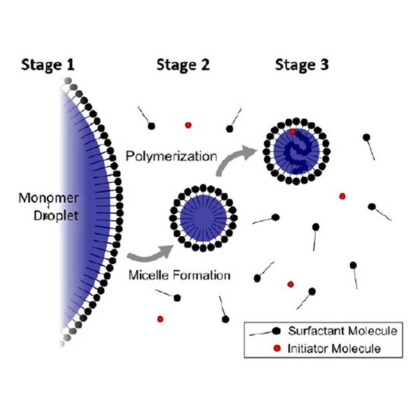
This note covers the history, theory and processes used for emulsion polymerization. It also examines the importance of measuring the concentration of latex nanoparticles produced by emulsion polymerization for a biotech application developed by the Gianneschi lab at UCSD. The Gianneschi case study includes data from HORIBA’s ViewSizer 3000.

Continue reading

Infections Titer Application Note

Biological
Applications
Infectious Titer ViewSizer 3000

Achieving Infectious Titer with Multi-laser Nanoparticle Tracking Analysis (NTA)

The viral vector market became highly active after the launch of a number of Advanced Therapy Medicinal Products (ATMPs). Based on the number of cases in clinical trials and its success rate to date of this writing, we can likely anticipate a cascade of FDA-approved products within years.

The complexity of viral particles remains one of the biggest hurdles in the development process. In this note, the upstream process of analyzing viral preparations is addressed, as well as the use of multi-laser nanoparticle tracking analysis (NTA) as a cost and time efficient method to measure size, count virus particles, and correlate to infectious titer.

Continue reading

Vaccine Manufacturing Application Note

Industrial
Applications
Vaccine Manufacuring
ViewSizer 3000

Particle Analysis in Vaccine Manufacturing and Development

One Size Doesn’t Fit All

Size matters in vaccine delivery systems. Nanoparticles smaller than 200 nm generally present a greater immunogenic response than micro-particles larger than 1 micron. This rather simple statement is based on the common understanding that particles with sizes resembling the dimensions of viruses are treated like viruses by the body. In the case of manufacturing for novel COVID-19 vaccine, adenovirus around the same size as SARS-Cov-2 (median of roughly 90-100 nanometers) are manipulated as carriers (or viral vectors) to trigger spike proteins production. In contrast, published literature showed that the effect of vaccines given orally, intranasally, or via other mucosal surfaces favor micro- over nanoparticulate formulations due to higher antigen load. The size of the impurities also significantly affects vaccine efficacy. In sum, many vaccine formulation ingredients should have controlled particulate size, size distribution, and count throughout the process of development, manufacturing, storage, and administration.

Vaccine Commercial Production

The upstream process of vaccine preparations requires careful virus characterization to achieve optimized infectivity and stability. Infectious titers are used to determine the concentration of viral particles that can transduce cells and its virus load in a sample. Two established analytical techniques to measure infectious titers are:

  • Viral Plaque Assay (VPA)
  • Quantitative Polymerase Chain Reaction (qPCR)

Both approaches quantify the amount of virus present in a solution. Viral Plaque Assay for lentivirus, for example, takes up to two weeks of incubation time to determine its result in a form of Plaque Forming Units (PFU) per mL. The counting of PFU is also subjective, resulting in low reproducibility from one analyst to another. qPCR, on the other hand, does not discriminate between whole, broken, empty, aggregates, infectious or non-infectious viruses; it merely determines relative viral gene expression and correlates the value back to PFU. The drawback is that the qPCR requires prior knowledge of the viral genome sequence, can be costly, and analyzes concentration of genomic material, not infectious virus since uncoated RNA or DNA may exist in a sample, but, without a coat, be unable to enter a cell.

Figure 1: Measurement result of a human viral vector sample. Note the distribution displayed presence of both host cell debris and aggregates.

Particle concentration analysis results from ViewSizer 3000™ multi-laser Nanoparticle Tracking Analysis (NTA) correlate with PFU, similar to qPCR. Due to three simultaneous operating lasers, it quantifies not only the intact viral particles but also infectious aggregates. Figure 1 demonstrates the measured size distribution of a sample of human viral vector, a virus candidate used in vaccine manufacturing. Note the significant population of larger particles.

Download Application Note 1: Achieving Infectious Titer with multi-laser Nanoparticle Tracking Analysis (NTA) on the right hand side

The ability to effectively analyze the entire size range allows the ViewSizer 3000 to successfully correlate known infectious titer to the total particle concentration with a R2 value of greater than 0.9, proving the multi-laser NTA technique a new, cost-effective and time-efficient alternative to VPA and qPCR.

Figure 2: Infectious titer correlation.

Viruses and Virus-Like-Particles (VLP)

Virus-like particles (VLP) are meant to mimic the virus of interest to provoke a therapeutic effect (such as immunity) without the expense of virus or risk of infection. They have been the focus of countless investigations on innovative vaccines. The size of VLPs is similar to that of viruses, which typically ranges from a few tens to a few hundred nanometers. Laser diffraction is an ensemble technique that allows quick, routine analysis. The technique is also fundamentally more sensitive to larger particles such as contaminants. Depending on how the VLP is manufactured, fragments of starting material will likely be present in the sample at larger sizes than the VLP itself. This arises when materials (such as emulsions) are prepared with a large or broad size distribution and then the size reduced to produce a final product. Remaining large particles can lead or unwanted immune response or issues with filtration in subsequent processing (such as filtration sterilization). To analyze viruses and VLP alike, it is essential a technique covers a wide dynamic particle range.

An example size result (below Figure 3) shows three separate populations. The finest (smallest particle size) population is the VLP itself. The remaining populations (with diameters over about 1 micron) are starting material that has not yet been sheared or otherwise milled to finer sizes. The LA-960 can report size metrics for the entire population as well as metrics for each individual population using the Multimodal Report.

Figure 3: Particle size distribution and results for a VLP material as measured by the LA-960. This sample shows three separate populations. The finest (smallest particle size) population is the VLP itself. The remaining populations (with diameters over about 1 micron) are starting material that has not yet been sheared or otherwise milled to finer sizes.

Exosomes for a New Generation Vaccine

A subgroup of extracellular vesicles (EV) known as exosomes play an increasingly important and intricate role in diagnosis and treatments of various diseases. They are responsible for transferring genetic material and cell-to-cell communication by carrying various nucleic acids, including RNA, lipids and proteins. Its immunogenic properties reportedly correlate with the amount of associated antigens, according to many published reports, thus, creating an opportunity for potential vaccine development.

Exosome particle size and concentration are especially scrutinized as they present important clinical information. To date, however, EV research still lacks standardization for its purification process. It also have been limited by the analytical technologies used to measure them. It is well understood, however, that EVs are a heterogeneous group of particles with a range of sizes and biogenesis; the size distribution are expected to be wide, even after processing.

The ViewSizer 3000 features three simultaneous operating lasers at 635 nm (red), 520 nm (green), 445 (blue) and a color camera to accurately visualize particles of large dynamic range. It overcomes the common drawback of conventional NTA, which is the failure to size particles accurately in a polydisperse sample. Human Preadipocyte (Mesenchymal Stem Cell) Exosomes (100 ug) samples acquired from ZenBio was measured using the ViewSizer 3000. The result was validated by a tunable resistive plug sensing technique and showed a profile with D50 particle diameter of 148 nm and total particle concentration of 5.7 x 10⁷ particles/mL.

Figure 4: Particle size and concentration distribution of exosome.

Download Application Note 2: Particle Size Distribution and Concentration of Exosomes

Conclusions

Viruses, VLP’s (such as adjuvants) and exosomes can be analyzed for size and concentration using a multi-laser nanoparticle tracking analysis (NTA) instrument, the ViewSizer 3000. Most samples show a wide size distribution that frustrates single laser NTA. If only size distribution is needed, such as in a size reduction process, laser diffraction has also proven useful.

Continue reading

Single and Multi-point comparison Application Note

Industrial
Applications
Single- and multi-point comparison

Comparison of Single-Point and Multi-Point Surface Area Measurements

The Flowing Gas Technique for determining BET Surface Area has been in use for over 70 years. Many facets of the technology make it a very attractive alternative to the Static-Volumetric approach.

First and foremost is the fact that the detection is done by measuring a gas concentration difference instead of an absolute pressure. Difference measurements are typically more accurate than many absolute measurements.

The speed of analysis and the resulting high sample throughput is also quite attractive. As a result, for routine QA/QC analysis, there has been a renewal of interest in this technique.

Continue reading

Surface Area Standards

Industrial
Applications
Low Specific Surface Area Standards
Horiba SA-9600

Measuring Low Specific Surface Area Standards with the SA-9600

A feasibility study to measure low specific surface areas with the SA-9600 Series was performed using Certified Reference Materials BCR 169, 170 and 172 from the European Commission Joint Research Centre. The reference materials consist of two alpha-alumina powders and one quartz powder, with certified values 0.1, 1.05, 2.56 m2/gram, respectively. The results show the SA-9600 can measure low specific surface area, with great agreement with certified values.

Continue reading

Metal Powder Application Note

Industrial
Applications
Metal Powders SA-9600

Metal Powder Properties: A Case with Low Specific Surface Area

Powder metallurgy is the study of transforming metal into powder and the consolidation of powder into the desired final product through methods such as sintering, compaction, blending, injection molding, or extrusion. Powder metallurgy is the essence of 3D additive manufacturing and it covers a broad spectrum of traditional applications including orthopedic implants, dental restorations, or paint pigments. The success of any powder metallurgical process, however, depends heavily on the understanding and control of the metal powder characteristics.

Continue reading

Food Packaging Application Note

Industrial
Applications
Food Packaging BeDensi T Pro

Optimizing Food Packaging Size by Measuring the Tapped Density

A reasonable packaging size in the food industry is important not only to ensure the success of the packaging process, but also to decrease the cost of transporting products. This application note explores how the size of the food powder container is determined by measuring the bulk density and tapped density. In this research, three types of protein powders, including whey protein, soy protein and whey-soy protein mixture, were analyzed by an automatic tapped density tester – The BeDensi T1 Pro. The result demonstrates that the instrument relies entirely on providing food manufacturers with reliable information to determine the optimum packing size and choose a container that is satisfying to the customer.

Continue reading

Pharmaceutical Application Note

Industrial
Applications
Pharmaceuticals BeDensi T Pro

How to Perform a Standardised Tapped Density Test for Pharmaceutical Powders

Tapped density is a significant parameter to explore the compressibility and flowability of pharmaceutical powders, which is useful to promote the approach of QbD and GMPs. Standardization of apparatus and procedure is vital to get meaningful repeatable results. In this application note, standardized tapped density tests of three excipients were performed by the BeDensi T3 Pro with 3 workstations. It is worthy of note that this highly efficient and economic tester is designed to meet the USP and EP standards fully.

Continue reading

Resins Application Note

Industrial Applications
Resins BeNano Series

Using BeNano 90 Zeta to measure the particle size and zeta potential of multicolor UV-sensitive resins

Multicolor UV-sensitive resins are widely used in the fields of 3D printing, inks, and paintings. When the size of the added particles is down to the nanoscale, many properties of the resin, such as dispersibility, uniformity, curing properties, glossiness, and brightness, will be improved greatly. However, the nanoparticles in resin are not always dispersed at the nanoscale as expected. BeNano 90 Zeta is a powerful tool for measuring the nanoparticle size and zeta potential to help in investigating the actual size of particles dispersed in resin and the dispersing stability.

Continue reading

Pesticides Application Note

Industrial
Applications
Pesticides Bettersizer ST

Application of Laser Particle Size System in Pesticide Industry

The particle size distribution of pesticides directly affects the trajectory of particle movement, surface energy and adhesion. This application note shows that the laser analyzer can not only optimize the component content in the formulation development, but also effectively monitor the particle size distribution of the pesticide in the process production to ensure the stability of product performance.

Continue reading

Gypsum Application Note

Industrial
Applications
Gypsum Bettersizer ST

Measuring Particle Size Distribution of Gypsum Using Laser Diffraction

The performance of gypsum such as setting time, compressive strength or density deeply relies on its particle size distribution. Bettersizer ST, an analyzer for quality control, allows the measurement of particle size distribution of gypsum. In this note, two gypsum samples were rapidly and accurately measured with Bettersizer ST. Outstanding repeatability was presented subsequently by measurements of a ground sample, which indicates its excellent reliability.

Continue reading

Abrasives Application Note

Industrial
Applications
Abrasives Bettersizer ST

Application of Laser Particle Size Analyzer in Quality Inspection of Silicon Carbide Abrasive Grains

The particle size distribution is one of the most important characteristics of abrasive grain products. In this application note, we will be looking at the particle size distribution of four batches of black silica carbide using a Bettersizer ST laser diffraction particle size analyzer. The stability of the production process can be evaluated by comparing the particle size distribution of abrasive grain products with each other. The particle size distribution of different abrasive grain products can be determined and be compared to ISO standards to determine if they are up to standard or not.

Continue reading

Ceramic Powders Application Note

Industrial
Applications
Ceramic Powder Bettersizer ST

Analysis of Particle Size Distribution of Ceramic Powder Based on Laser Diffraction

During ceramic powder processing, the particle size distributions of powder, slurry and granule are different, which are necessary to be monitored. In this note, three forms of aluminium oxide, namely powder, slurry and granule were measured with the Bettersizer ST. And outstanding repeatability was demonstrated through the measurement of a granule sample.

Continue reading

Calcium Carbonate Powders Application Note

Industrial
Applications
Calcium Carbonate Bettersizer ST

Measuring Particle Size Distribution of Calcium Carbonate Powders with Laser Diffraction Method

Over a wide range of industries, different functions require different particle size distributions of ground calcium carbonate powders. That is the reason why particle sizing is a vital step for quality control of ground calcium carbonate. In this investigation, particle size distributions of three different ground calcium carbonates were measured by the laser diffraction method. Typical size values and size distribution curves were compared to evaluate the quality and stability of the sizing process of the three samples.

Continue reading

Pesticides Application Note

Industrial
Applications
Pesticides Bettersizer ST

Inspect the Quality of Pesticides with Laser Diffraction Particle Size Analyzer

From the key active ingredients to the final pesticide product, particle size is known to have an important role in the residual period, the biological activity, and the stability properties of pesticides. For this reason, measuring the particle size distribution plays a vital role in determining the quality of pesticides during QC inspection. In this application note, particle size distributions of suspension concentrate samples were measured by the laser diffraction method. Typical size values and size distribution curves were compared to evaluate the quality of pesticide samples and to help to optimize the milling process and produce a pesticide that is fit for the specific application.

Continue reading

Lithium-ion Battery Application Note

Industrial
Applications
Lithium-ion Battery Bettersizer ST

Improving the Tapped Density of the Cathode Material to make a Lithium-ion Battery Hold More Energy

Tapped density is one of two important physical properties of electrode materials and affects the energy density of a Li-ion battery (LIB). The other important physical property is the particle size distribution which provides the appropriate information to optimize the grinding parameters during production. Improving the tapped density can also optimize the high- energy-density during LIB manufacture. Thus, it is necessary for the LIB producer to determine in advance, what is the most optimal and achievable tapped density and then using this parameter as the “gold standard” measure samples from the process during production until they match or come close to the “gold standard” measurement. The easy-to-use BeDensi T Pro series is an ideal tapped density tester because it is an economical device which delivers an exceptional performance with no compromises.

Continue reading

Chinese Medicine Powder Application Note

Industrial
Applications
Chinese Medicine Bettersizer 2600

Research on Particle Size Measurement of Chinese Medicine Powder by Laser Particle Size Analyser

The particle size and particle size distribution, which are related to the quality of the products and the safety of the drugs, are the important physical properties of the Chinese medicine powder. However, because of the irregularity and inhomogeneity of particle size, the results obtained by different measurement methods are different. Laser particle size measurement has been widely used in the determination of traditional Chinese medicine by its fast operation, wide measuring range, and good reproducibility. This paper focused on the principles and characteristics of laser particle size measurement and its application of Chinese medicine powder.

Continue reading

Domperidone API Application Note

Industrial
Applications
Domperidone Bettersizer 2600

Research on Particle Sizing Dispersion Methods of Domperidone API by Laser Particle Size Analyser

For particle size distribution measurement of Domperidone API, both wet and dry dispersion methods could provide high precision results. However, since Domperidone API is fragile, adequate data support is required for sample dispersion, especially for dry dispersion method.

In this application note, compared with dry dispersion, wet dispersion was observed to provide data with better repeatability, correlation, and rationality. Therefore, wet dispersion method is relatively reasonable to analyse particle size distribution of Domperidone API.

Continue reading

Lactose Application Note

Industrial
Applications
Lactose Bettersizer 2600

Research on Particle Size Measurement of Lactose by Laser Particle Size Analyser

Lactose is one of the most common kinds of tablet excipients. The USP has clear regulations on raw materials particle sizing by laser diffraction method, such as the structure and principle of the instrument, the specific method of dry and wet dispersions, the factors in the measurement process, etc. However, for specific lactose, there is no related instruction on how to choose the dispersive pressure and how to evaluate the results of dry and wet methods. This note carried out a systematic research on lactose particle size distribution measurement in accordance with the USP and the ISO 13320.

Continue reading

Coffee Application Note

Industrial
Applications
Coffee Bettersizer 2600

Application of Laser Particle Size Analyzer in Coffee

This note explores the effects of coffee grinding method, particle size and particle size distribution on coffee quality, and introduces the measuring methods and principles for optimizing coffee particle size and particle size distribution. The study results show that the laser particle size analyzer can effectively analyze and monitor the particle size and particle size distribution of the coffee in the grinding process, ensuring the homogeneity of the product and improving the quality of the coffee and also contributing to quality control in the development and production of coffee grinding equipment.

Continue reading

Cement Application Note

Industrial
Applications
Cement Bettersizer 2600

Rapid Laboratory Particle Size Analysis of Cements Using Laser Diffraction

The large power demands of finish milling mean that improved monitoring of the grinding efficiency and optimization of the classifier speed yields an in-specification product with significant energy efficiency improvements and ultimately cost savings. This is best achieved by having a laser diffraction which is quick and easy to use with consistent repeatable results being attained no matter which operator is using the system. In addition, by having control standard results for each cement grade maintained inside the computer database, all newly produced cement for all grades can be compared in seconds to the ideal products fineness parameters. The Bettersizer 2600 has all this functionality in its software and provides the rapid laboratory fineness analysis to prove the cement meets the specifications and is thus fit for purpose.

Continue reading

Milk Powder Application Note

Industrial
Applications
Powdered Milk Bettersizer 2600

Particle Size Analysis of Milk Powder by Laser Diffraction

In the production and application process of milk powder, the particle size of milk powder directly affects its final taste, sensory properties and quality characteristics. The Bettersizer 2600 can be used by milk powder manufacturers or relevant equipment manufacturers to accurately monitor the size changes of milk powder during production, packaging, storage and application process, as well as to better understand the relationship between dairy product formulation and quality.

Continue reading

Pharmaceutical Application Note

Industrial
Applications
Pharmaceutical Bettersizer 2600

Particle Sizing with Dry Dispersion Can Be the Best Choice for Pharmaceutical Analysis

This paper looks at laser diffraction technology which when initially conceived only measured particle size by diluting a sample with a suitable diluent and pumped the mix through a sample measuring cell. It explains how the move away from using solvents encouraged the use of measuring sample dry rather than wet and explains what the barriers to dry measurement were and how they were overcome during a period of 25 years.

Pharmaceutical powders are generally considered to be some of the most cohesive a particle scientist will come across and generally are very demanding when using the wet method. In this paper we will demonstrate how a modern dry laser diffraction is able to analyse these cohesive samples and achieve reliable repeatable results with an eco-friendlier method.

Continue reading

Mineral Pigment Application Note

Industrial
Applications
Mineral Pigment Bettersizer 2600

Particle Size of Mineral Pigment and Colour Hiding Power

Particle size variation in mineral pigments leads to different hiding power and diverse chromatic gradation, attributed to the light scattering effect. The Bettersizer 2600 can provide meticulous measurement of granularity and support in finding the optimal balance between particle size and desired hiding power, ensuring consistency in pigment and coating products.

Continue reading

Lithium Battery Cathode Materials

Industrial
Applications
Battery Bettersizer 2600

Application of Laser Particle Size Analyser in Lithium Battery Cathode Materials

In order to achieve high energy storage, stability and safety performance, it is necessary to strictly control the particle size distributions of lithium battery cathode materials. Therefore, it is important for battery manufacturers to measure the particle size distribution of electrode materials quickly and easily, with the Bettersizer 2600 laser particle size analyser.

Continue reading

Ceramic Powders Application Note

Industrial
Applications
Ceramic Powder Bettersizer 2600

Application of Laser Particle Size Analyser in Ceramic Powders

Accurate measurement of the particle size distribution of ceramic powders is extremely necessary in the production of modern ceramic components. It has been proved that the particle size and dispersibility of a ceramic powder can be determined by the Bettersizer 2600, and the test results have a high repeatability.

Continue reading

Chocolate Application Note

Industrial
Applications
Chocolate Bettersizer 2600

Particle Size Analyzing of Chocolate by Laser Diffraction

The manufacturing process and final characteristics of chocolate are significantly affected by particle size in many ways. As less production costs and better chocolate quality are desirable, only with the help of high-performance laser diffraction instruments, manufacturers are able to control particle size distribution of intermediates as well as final products in chocolate production in a highly efficient way. In this note, the measurements of chocolates of different types (dark, milk, white) from various countries were successfully performed by the Bettersizer 2600, and the particle size changes were displayed.

Continue reading

Coffee Application Note

Industrial
Applications
Coffee Bettersizer 2600

Investigating the Relation Between Coffee Extraction and Ground Coffee Size

Particle size and size distributions of ground coffee significantly affect the extraction level and the flavor quality of brewed coffee. Monitoring the particle sizes and size distributions in ground coffees is necessary. In this note, different ground coffees were successfully characterized by the Bettersizer 2600, which is a sophisticated and reliable instrument that can provide particle sizing solutions to the coffee industries.

Continue reading

Pigments Application Note

Industrial
Applications
Pigments Bettersizer 2600

Application of Laser Particle Size Analyser in Pigments

Coarse particles influence the color of pigments, and particle aggregation that occurred during storage reduces the stability of product performance. The Bettersizer 2600 enables the manufacturers to monitor the particle size and its distribution of pigments in the production and storage process. The instrument’s wide detection range and high resolution allow all pigments to be measured accurately, and ensure excellent batch-to-batch reproducibility.

Continue reading

Abrasives Application Note

Industrial
Applications
Differing Abrasives

Application of Image Particle Size & Shape Analysis System in Abrasive

This application note compares the particle size and particle shape distribution of different kinds of abrasive by laser scattering method and image method. The results show that the accuracy of particle size measurement results adopted by image method is better and coarser particle resolution is higher, which can effectively monitor the particle size, particle size distribution and ovality, circularity in the abrasive production process to ensure the uniformity of the product, thus improving the performance of the abrasive tool, therefore, the image method is one of the indispensable detection methods in the abrasive industry.

Continue reading

3D Printing Application Note

Industrial
Applications
3D Printing

Particle characterization for additive manufacturing: Analysis of the key parameters particle size and shape using only one instrument

Additive manufacturing (AM) also known as 3D printing, is taking off to produce a wide range of components more efficiently, sustainably, and cost-effectively. In order to achieve success, it is essential to accurately characterize raw materials, such as metal powders and polymer powders. To meet this need, Bettersizer S3 Plus offers characterization solutions. In this app note, we explained the importance of particle size and shape characterization for raw materials and analyze the key parameters using only one instrument.

Continue reading

Abrasives Application Note

Industrial
Applications
Abrasives

Combining Laser Diffraction with Dynamic Image Analysis to Improve the Characterization of Abrasives

Size is not enough to ensure the consistency of abrasives and it is well known within the abrasives industry that the shape of the particles is equally as significant a parameter to control. The Bettersizer S3 Plus has proven to be able to characterize the size and shape of the abrasives simultaneously and provide much more information than conventional laser diffraction analyzers.

Continue reading

Soils and Sediments Application Note

Industrial
Applications
Soils and Sediments

Exploring Size and Shape on Soils and Sediments of the Moon, the Earth and the Ocean

Soil and sediment analysis is essential for human beings, which provides fingerprints to their origin. The main categories of soil and sediment analysis include hydrology and geology studies. Particle size and shape are challenging in soil and sediment analysis. Why? Soil samples are polymorphic and always cover a wide size distribution range. The Bettersizer S3 Plus analyzes the particle size over a wide range from 0.01 μm to 3.5 mm, fully meeting the needs of soil and sediment size measurements. This application note will focus on three different applications covering lunar regolith, desert, and marine sediments and explore the differences of particle size and shapes in three samples.

Continue reading

Lithium-Ion Battery Application Note

Industrial
Applications
Lithium-Ion Batteries

Investigating the Particle Size and Shape Influences on Anode Energy Density of Lithium-Ion Batteries

The lithium-ion batteries (LIBs) have been widely used in
variety of applications due to its advantages of long storage
life, no memory effect, and low self-discharge rate. With the
rapid increasing demands of LIBs in electrical products, the
production of higher energy-density batteries has attained
manufacturers’ attention because of the needs of storing
more energy.
The energy density of the anode can be significantly
improved by optimizing the particle size and shape of the
graphite

Continue reading

Ceramic Agglomerate Application Note

Industrial
Applications
Ceramic Aglomerates

Particle Size Measurement and Agglomerates Detection of Ceramic Materials During Production Process

Accurate measurement of ceramic powders is crucial in the ceramic manufacturing. The Bettersizer S3 Plus has been proven to accurately measure the particle size and size distribution, and effectively monitor the agglomeration existing in ceramic powder materials. Therefore, the Bettersizer S3 Plus is a valuable tool to display both particle size and shape results. With the assistance of the Bettersizer S3 Plus, manufacturers are able to produce high performance ceramic products.

Continue reading

Powder Coatings Application Note

Industrial
Applications
Powder Coatings

Particle Size and Shape Analysis of Powder Coatings

In the production process of powder coatings, particle size is one of the most important physical properties, which not only affects the spraying performance of finished coatings, but is also closely related to the entire production process of coatings. In this note, different kinds of powder coatings have been successfully characterized by laser diffraction analysers, which have replaced conventional methods to a large extent mainly due to the advantages of the technology including ease of use, fast operation and high reproducibility, which are powerful tools for the powder coating industry.

Continue reading

Ceramic Products Application Note

Industrial
Applications
Ceramic Products

The Quality Control of Advanced Ceramic Products by the Bettersizer S3 Plus

The global demand for advanced ceramics, with the unique thermal, wear, and corrosion resistant capabilities, in biomedical, aerospace industry, precision tools, electronics, and environmental fields is on the increase. Optimizing and controlling the particle size distribution of powder to improve the microstructure of ceramic products are crucial to the final performances. The Bettersizer S3 Plus and BT-A60 autosampler can provide ceramic powder producers and ceramic product manufacturers with a highly automatic and time-saving method for measuring large numbers of samples. The high performances and the combination of dynamic image analysis enable the Bettersizer S3 Plus to be a reliable and powerful tool for quality control during any process of ceramic production.

Continue reading

Soy Milk Application Note

Industrial
Applications
Soy Milk

Particle Size Analysis: Exploring the Impact of Homogenization on Soy Milk

To enhance the taste and stability of soy milk, homogenization, subjecting the liquid to intense shearing, breaking down large fat globules particle and protein clusters, is a crucial step in the manufacturing process. Bettersize can provide the soy milk particle analysis with the combination of two instruments: Bettersizer S3 Plus and BeVision S1, so as to ensure the overall quality of soy milk product and create a homogeneous liquid that effectively prevents fat floating and protein settling.

Continue reading

nCS2 Industrial Nanoparticles Application Note

Biological
Applications
Industrial Nanoparticles

Applications
Nanoparticle Analysis of Any Material

Spectradyne’s microfluidic technology enables detection of any nanoparticle type!

Overview

The Spectradyne nCS2TM and ARCTM employ a novel implementation of resistive pulse sensing to count and size nanoparticles quickly and with high resolution. Sizing precision of ±3% is achieved, with rates up to 10,000 particles/s. Because particles are measured electrically, not optically, particles composed of any material can be analysed, without a materials-influenced bias. In Spectradyne’s ARC, this individual particle sizing and counting capability is combined with individual particle fluorescence detection.

“The nCS1 is fast, accurate, and easy to use for routine particle quantification.”

– Hok Hei Tam, Flagship Labs 62 Inc

Materials

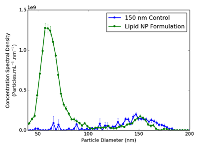
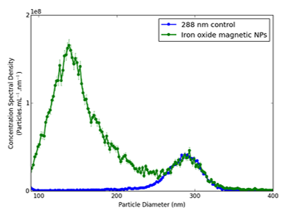
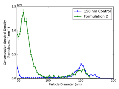
Nanoparticles are used in a broad range of applications. Metal-oxide nanoparticles such as TiO2 are used as additives in cosmetics (UV absorbers in sunscreen) and food (whiteners in frosting). Semiconductor nanoparticles are used for their unique optical properties (light-emitting quantum dots in digital displays). Gold nanoparticles are used in drug delivery and in-vivo targeted treatments. The nCS2 and ARC are able to accurately measure particles of any material. For example, here we show data where the nCS1 accurately measures the size and concentration of different types of nanoparticle. In each case results are shown with a measurement of the pure control particles (no additional particles) for comparison.

BioLector Microbioreactor

Continue reading

nCS2 Serum Application Note

Biological
Applications
Serum

Applications
Measuring nanoparticles in blood serum

Spectradyne’s technology allows the measurement of nanoparticles in serum!

Overview

The Spectradyne nCS2TM and ARCTM particle size analyzers can be used to analyze nanoparticles in blood serum, blood plasma, as well as in other weakly conducting analytes such as PBS or other salt-containing solutions.

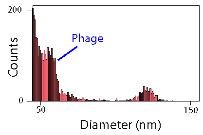
Below we display data showing the positive detection of bacteriophage in murine blood plasma. Left plot shows the raw particle detection data, of T7 phage in murine plasma. Center plot shows the histogram of effective particle diameters, showing a peak in particle concentration at 55 nm attributed to the bacteriophage. Left plot shows the concentration spectral density (CSD) of the blank plasma (blue dashed), phage-infected plasma (green dot-dashed) and the difference (red solid). These data also yield a 55 nm diameter for the phage. Integration of the peak gives the measured phage concentration of 5.3 × 10¹⁰ particles per ml, near that obtained by biological titre, of 1.5 × 10¹⁰ pfu/ml).

BioLector Microbioreactor

Continue reading

nCS2 Gene Therapy Application Note

Biological
Applications
Gene Therapy

Applications
Analysis of Gene Therapy Vectors and Nanomedicines

Spectradyne’s microfluidic technology has powerful applications in nanomedicine

Overview

Accurate quantification of gene therapy vectors and nanomedicines is critical at all stages of research and product development, whether for characterizing a production step or obtaining an accurate titer before assessing bioactivity. Spectradyne’s nCS2TM and ARCTM quantify all types of nanoparticle-based therapeutics quickly and accurately, and require only 3 microliters of the sample per measurement. The ARC instrument adds a fluorescent pheontyping capability to the particle size and concentration provided by Spectradyne’s nCS2.

Quantifying a virus-based drug product

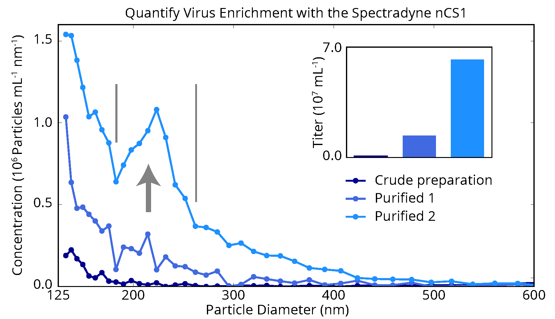
The figure below shows nCS2TM measurements of a proprietary virus-based drug product as it progresses through three stages of purification. While each stage of purification increases the concentration of particles over a broad size range, a clear enrichment of virus product is obtained with the second stage. These measurements provided the manufacturer of this product with a highly detailed picture of the purification process, and insights for optimization that could not have been obtained with any other method.

Gene therapy vectors

Because the nCS2 uses an electrical method, not light scattering, to count and size nanoparticles, it measures all particle types equally well. As a result, researchers count on the nCS2 every day to measure diverse range of gene therapy vectors and other nanomedicines, including:

  • Lentivirus, retrovirus, HSV and others
  • Lipid nanoparticles (LNPs)
  • Liposomes
  • Polymeric nanoparticles
  • Milled drug particles (API crystals)

Fluorescent phenotyping

Spectradyne’s ARC combines the microfluidic resistive pulse sensing of the nCS1 with a particle-by-particle fluorescence detection, allowing phenotyping combined with particle size and concentration measurements. The applications of this fluorescence-extended MRPS instrument allows the ARC to measure a diverse range of nanomedicine targets, including:

  • Polymeric nanoparticles
  • Milled drug particles (API crystals)
  • Lentivirus, retrovirus, HSV and others
  • Lipid nanoparticles (LNPs)
  • Liposomes
BioLector Microbioreactor

Spectradyne’s nCS2 enables monitoring virus purification stages

A virus-based drug product measured by the nCS2 after various stages of purification. Enrichment of the virus is clearly observed after the second purification step, and could not be measured so directly and practically using any other technology. Such high-resolution and quantitative measurements as these cannot be obtained with other technology, and enable more effective analysis of process parameters at all stages of the drug development process.

Continue reading

nCS2 Protein Aggregation Application Note

Biological
Applications
Protein Aggregation

Applications
Nanoparticle analysis of protein aggregation

Spectradyne’s microfluidic technology enables detection of any nanoparticle type!

The Spectradyne nCS1TM employs a novel implementation of the resistive pulse sensing method (MRPS) to count and size nanoparticles quickly and with high resolution. Sizing precision of ±3% is typically achieved, with measurement rates up to 10,000 particles/s.

“Submicron protein particle characterization by Resistive Pulse Sensing results in high size resolution and provides an estimate of particle counts, thereby, provides better insight into particle size distribution compared to the traditional light scattering techniques.”
G.V. Barnett, J.M. Perhacs, T.K. Das and S.R. Kar, Submicron Protein Particle Characterization using Resistive Pulse Sensing and Conventional Light Scattering Based Approaches in Pharm Res. 35, 58 (2018) doi: 10.1007/s11095-017-2306-0
A publication from Bristol-Myers Squibb

Protein aggregation

Spectradyne’s nCS1 delivers two significant advantages for detecting aggregates in protein solutions: First, individual particle measurements (no ensemble-averaging) deliver accurate size distributions in highly polydisperse samples such as these. Second, the non-optical electrical detectionmethod ensures that protein aggregates, which are low-index contrast materials, are accurately represented in the distribution.

Particulates in parenteral drug development and production have always been a serious issue. In biologics, the issue is compounded by reported impacts of aggregates and particles on the product’s efficacy, safety and immunogenicity. FDA regulations strongly recommend in-depth characterization of the identity and quantity of particles in protein therapeutics.

While regulations require measurement of larger particles (>1 μm), it is desirable to detect and characterize protein aggregates long before they are that large. Crucial decisions about formulation, processing, storage conditions, etc. must be made with an eye towards minimizing protein aggregation throughout the drug life-cycle.

Spectradyne’s nCS2TM

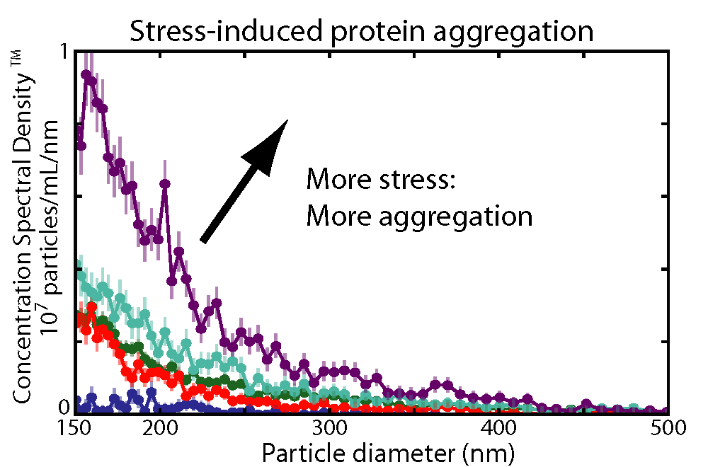
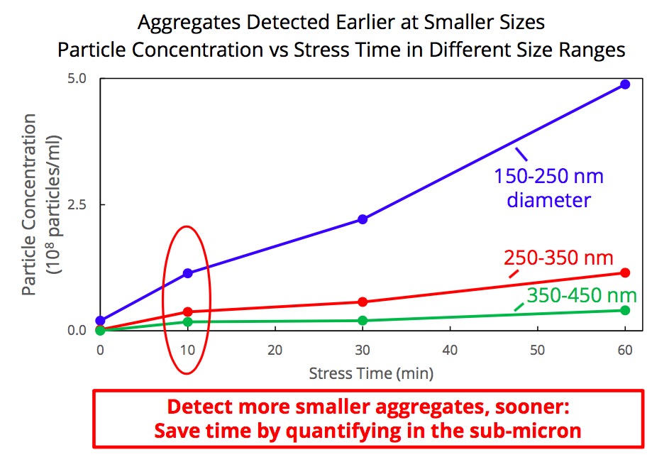
Spectradyne’s nCS2 is ideally suited for accurate quantification of submicron protein aggregates. The figure below demonstrates the power of the nCS2 to quantify protein aggregation in real world samples. Five formulations that had been stressed to varying degrees (0, 10, 20, 30, and 60 minutes) were provided by a large biopharmaceutical customer and analyzed “as received” on the nCS2 – no dilution or additives were required. The range of concentrations measured by the nCS1 spanned over four orders of magnitude in this instance. The quantitative results clearly show that increased stress causes more aggregates in this formulation.

Spectradyne’s ARCTM

Spectradyne’s ARC combines the MRPS functionality of the nCS2 with a fluorescence detection capability, allowing the fast and rapid detection of fluorescent tags combined with the accurate sizing and concentration measurements provided by microfluidic resistive pulse sensing (MRPS). This adds an extra dimension to your nanoparticle and protein aggregate detection capability.

BioLector Microbioreactor

Spectradyne’s nCS2 enables earlier detection of smaller aggregates

BioLector Microbioreactor

Continue reading

nCS2 Virology Application Note

Biological
Applications
Virology

Applications
Virus Quantification using Spectradyne’s nCS2TM and ARCTM

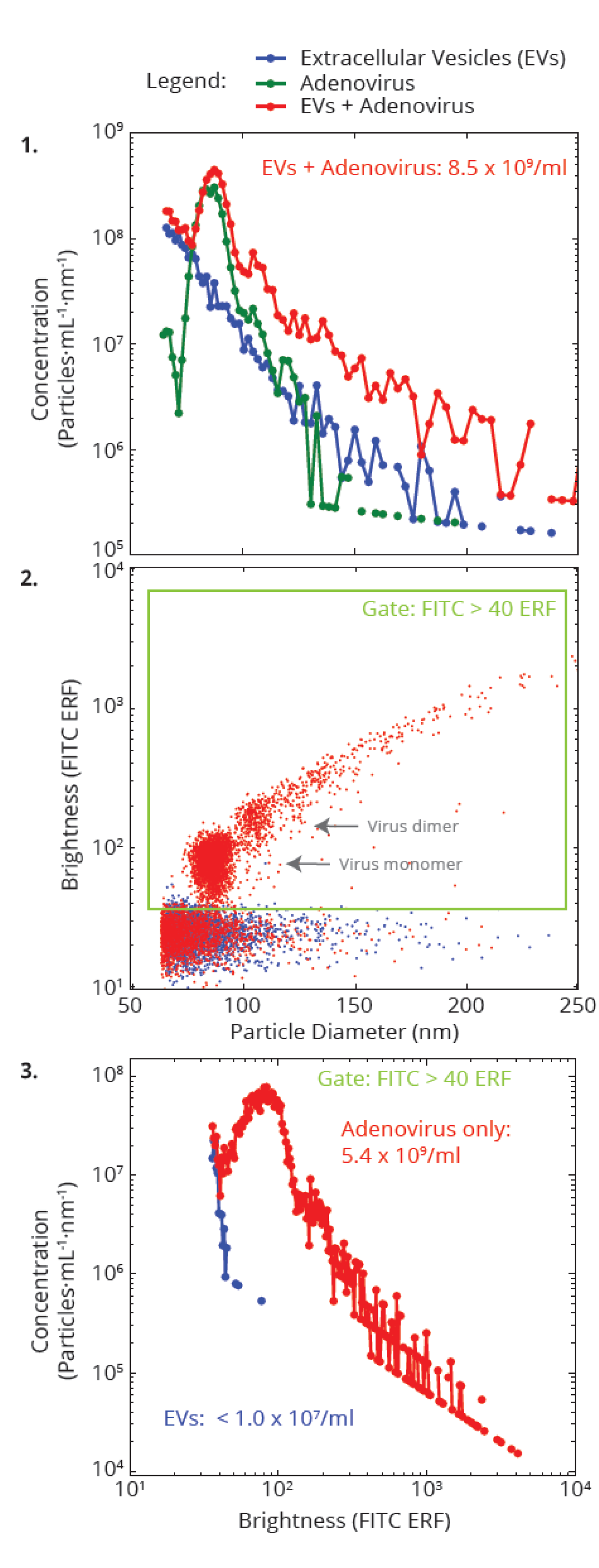
We’ve used the ARC to quantitatively measure the fluorescent labeling of adenovirus, using the ARCTM particle analyzer. These measurements were performed in a complex heterogenous sample, as described below.

The figure shows simultaneous particle sizing using MRPS and quantitative measurements of fluorescence, measurements made of EVs and adenovirus both together and in isolation. These data, which are discussed in more detail in this application note, demonstrate that the ARC quantitatively measures nanoparticle size, concentration, and internal payload fluorescence for virus and nanomedicine applications, and accurately analyzes fluorescent subpopulations, even in complex, heterogeneous samples.

BioLector Microbioreactor

Accurate Viral Titer in Minutes

Conventional methods for quantifying virus such as live biological titer can take days to return results. The modern fast pace of research and current examples such as COVID-19 necessitate faster measurements. Researchers in many areas of virus research including basic virology, gene therapy, and epidemiology are looking for more modern approaches for measuring virus.

Spectradyne’s nCS1TM and the fluorescent-detecting ARCTM deliver accurate viral particle counts in just a few minutes using microfluidic resistive pulse sensing (MRPSTM), which in the ARC is coupling with particle-by-particle fluorescence detection. The technology is fast, easy to adopt and employs disposable microfluidic cartridges for analysis — and it only requires a few microliters of your precious sample for analysis.

nCS2TM key features for virus quantification:

Total virus particle count in minutes

Disposable analysis cartridges — no laborious cleaning

Only uses 3 microliters sample required

The ARCTM adds individual particle fluorescence, with fluorescence quantified using NIST-traceable standards.

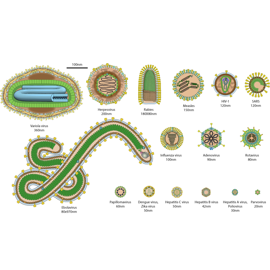
How big is your virus?

Virus come in a large range of different sizes and shapes. Some, such as the hepatitis and parvovirus, are roughly spherical and less than 50nm in diameter; some, such as the virus that causes Ebola, are long flexible cylinders with lengths as large as 1 μm; and some are spherical and of intermediate sizes, in the range of 75-300 nm in diameter.

In the figure below, we display a chart showing the size and shape of a range of different virus (chart obtained from Expasy).

BioLector Microbioreactor

Spectradyne’s nCS2TM can measure particles such as virus down to diameters less than 50nm and up to 10 μm in diameter. Contact us today if you’re interested in finding out more.

“We eagerly purchased Spectradyne’s current MRPS technology for the value it delivers in therapeutic retroviral quantification. We have been impressed with the results it delivers and how practically it fits with our industrial process flow…”

Better science

For measuring the infectivity of viruses, testing the immunogenicity of different viral preparations, and almost any down-stream analysis, accurate virus concentration measurements are critical for performing well-controlled experiments.

A retroviral drug product was serially diluted and the concentration measured as a function of size showing the excellent concentration linearity of the nCS2TM:

Serial dilutions of a retroviral drug product accurately quantified by Spectradyne’s nCS1TM.

More and more researchers are choosing the nCS2TM for virus quantification because it is fast and easy to use and delivers a more accurate analysis of their virus samples than they can obtain using any other method.

Continue reading

Liposomes and Lipid Nanoparticles Application Note

Biological
Applications
Liposomes and Lipid Nanoparticles

Liposomes and Lipid Nanoparticles

Liposomes and lipid nanoparticles (LNPs) are very similar in basic physical structure. These are both used as drug delivery vehicles in the body. Traditional liposomes have a lipid bilayer surrounding an aqueous pocket, while LNPs typically only have a single phospholipid outer layer encapsulating the interior, which can be non-aqueous.

As carriers for drug products (DPs), these both provide distinct advantages in that the exterior layer protects the DP from external degradation, and the physiochemical properties of these particles can be optimized for better specificity in the location and rate of drug delivery. For example, techniques such as pegylation can be used with LNPs to boost stability and increase circulation time in order to deliver more efficiently to targeted sites within the body. There is tremendous growth in the use of liposomes and LNPs as delivery vehicles for highly specialized drug payloads, such as mRNA therapies: Both of the initial SARS-CoV-2 vaccines approved for use (BioNTech/Pfizer and Moderna) use LNPs as delivery mechanisms for mRNA DPs.

Accurate Liposome and LNP quantification

One of the distinct advantages of using liposomes or LNPs is that both types of particles can be manufactured with very narrow, uniform particle size distributions. The actual size of the DP delivery envelope can be a critical part of the targeting mechanism for these drugs. Different size particles can target specific organs or tissue types, and, if small enough, can cross the blood-brain barrier to deliver directly to the brain.

This means that the ability to precisely measure particle size distributions for liposomes and LNPs is of critical importance. Spectradyne’s nCS1TM delivers higher-resolution measurement of particle size when compared to optical techniques like dynamic light scattering (DLS) and nanoparticle tracking. At the same time, it also delivers industry-leading concentration measurements, which are also critical because concentration correlates directly to dosage.

Spectradyne’s ARCTM combines the microfluidic resistive pulse sensing of the nCS1 with a particle-by-particle fluorescence detection, allowing phenotyping combined with particle size and concentration measurements. This allows the identification of liposomes through their particle-by=particle fluorescence characterization combined with size and concentration analysis.

Liposomes

In the plot above, you can see two LNP samples measured using Spectradyne’s nCS1 and also characterized using dynamic light scattering (DLS). DLS was unable to distinguish between the two formulations, which clearly have different mode sizes, and quite different overall distributions. The actual difference detected by Spectradyne’s nCS1 for mode size could affect the ability of the final DP to reach its intended target, so knowing this value exactly is critical for many applications.

Liposomes

In the second plot above, we are interested in the repeatability of the manufacturing process. The three samples represent three different production cycles for LNPs synthesized using identical recipes and process conditions. The data, taken using Spectradyne’s nCS1, show the three production replicates are very similar in particle size distribution and overall concentration. The DLS data is, by contrast, not usable, as the high bias of light scattering to larger-size particles totally misrepresents the actual sizes, and the high PDIs imply variability that doesn’t actually exist!

The lesson to learn from this: To get the most accurate particle size and concentration methods for your liposomal and LNP-based formulations, you need to use Spectradyne’s nCS1 MRPS system!

Continue reading

Extracellular Vesicles and Exosomes Applications Note

Biological
Applications
Extracellular Vesicles and Exosomes

Extracellular Vesicles and Exosomes

Extracellular vesicles (EVs) are biological particles ranging in size from about 30 – 10,000 nanometers in diameter. Extracellular vesicles comprise diverse types of particles having a broad range of physical properties and biological origin, and include exosomes, ectosomes, microvesicles, microparticles, oncosomes, and apoptotic bodies.

Extracellular vesicles have roles associated with an equally broad set of biological processes, including immune response, transfer of functional proteins and nucleic acid, elimination of unwanted materials, nutrition, surface-receptor mediated cell signaling, and numerous conditions of disease including cancer metastasis. Because of their involvement in so many physiological processes, extracellular vesicles hold enormous potential as therapeutics and biomarkers of health and disease. In these applications, accurate quantification of extracellular vesicles is critical for performing well controlled science, as well as ensuring safety and efficacy of extracellular vesicle-based products.

Accurate quantification with MRPS and simultaneous single-particle fluorescence

Spectradyne’s Arc delivers single-particle fluorescence combined with MRPS measurements, yielding size and phenotype for each individual nanoparticle, providing unparalleled individualized characterization not available with any other instrument. The Arc is fast and easy to use, and requires only 3 microliters of your sample to help you do better EV science.

Subpopulation analysis with fluorescence

Almost all biological nanoparticle samples contain heterogeneous populations of many different particle types. ARC can measure the size and concentration of the entire distribution using MRPS, and simultaneously measure and display only those particles in the sample that are positive for fluorescence in up to 3 different channels.

EV's 3 ways

The example above shows that for an EV sample containing 1.26E+10 particles/ml overall, only 1.03E+09 particles/ml, or 8.1%, are tagged FITC- positive. In the same way, one can easily measure subpopulations in a virus sample, for instance.

Accurate quantification with microfluidic resistive pulse sensing (MRPS)

Spectradyne’s nCS1 delivers the most accurate concentration and size measurements of extracellular vesicles available in a practical bench top solution. The nCS1 is fast and easy to use, and requires only 3 microliters of your sample to help you do better EV science.

“The nCS1 is fast, accurate, and easy to use for routine EV quantification. We looked at other nanoparticle measurement solutions and the nCS1 is the only instrument that could measure the size of every nanoparticle of interest in as little as 3 uL of volume. Moreover, their customer service is superb and have went out of their way to ensure our nCS1 is always functioning well.”

-Joseph Sedlak, Co-Founder at Mercy BioAnalytics

Accurate concentrations yield better science

For measuring the biological activity of exosomes, characterizing vesicles of different biological origin and exploring vesicle biomarker applications, accurate concentration measurements are critical for performing well-controlled experiments.

The figure below shows a comparison of Spectradyne’s nCS1 to NTA and the gold standard, Cryo-TEM. The nCS1 measurements are in excellent agreement with those of TEM, and indicate that the exosome concentration increases to smaller particle size down to 50 nm. In contrast, NTA reports misleading results: A loss of counting efficiency is apparent for particles as large as 200 nm in diameter, leading to a 10,000-fold error by 65 nm. Critically, NTA reports a prominent peak in the exosome size distribution near 130 nm diameter that does not in fact exist. Read more about why this happens.

EV's 3 ways

More researchers are choosing Spectradyne’s instruments for extracellular vesicle quantification because these are fast and easy to use and deliver a more accurate analysis of their vesicles than they can obtain using any other method.

Continue reading

Therapeutics (Other) Application Note

Industrial and Biological
Applications
Other Therapeutics

Particle Characterisation in Biotherapeutics

Particle content is a critical quality attribute for many biologics that must be monitored to meet regulatory requirements like USP <788> and mitigate product safety risks.

Flow imaging microscopy is an established technique recommended by USP <1788> for subvisible and submicron particle analysis, revealing particle count, size, and shape, indicating their type and source.

Monitor the aggregation of adjuvants, nano-drug delivery systems, and other small particles to larger, potentially concerning, submicron and subvisible particles.

Use FlowCam to:

  • Detect aggregation and agglomeration of the active pharmaceutical ingredient (API) and drug delivery vehicles to improve product stability
  • Obtain images and particle morphology information not obtainable from orthogonal techniques to assess product degradation
  • Differentiate between inherent particles, intrinsic particles like glass flakes and silicone oil droplets, and extrinsic contaminants
  • Optimize and control particulates in your formulation

FlowCam – A Flexible Particle Analysis Solution

Apply flow imaging microscopy techniques to therapeutics including aggregates of drug delivery systems like liposomes, exosomes, and gold nanoparticles, and vaccine components like virus-like particles and adjuvants.

FlowCam is ideally suited to analyze samples containing larger particles like CHO cells, cell cultures and associated particles like Dynabeads™ and Tentagel™ beads, and hydrogel spheres.

Obtain size and morphology information of these particles that is related to product quality issues such as cell viability, misshapen drug delivery vehicles, and the form of any aggregates present.

  • Improving vaccine formulations by monitoring API and adjuvant aggregation
  • Characterising large liposome, exosome, and other drug delivery platform morphology with an automated microscopy technique
  • Optimizing cell concentrations and viability during biotherapeutic manufacturing
  • Observing Dynabead binding and measuring unbound bead concentrations in cell culture applications
  • Product

  • Industry

Continue reading

Protein Therapeutics Application Note

Biological
Applications
Protein Therapeutics

FlowCam for Protein Therapeutics Development and Manufacturing

Discover a high-throughput flow imaging microscopy platform for characterizing API aggregates and other particulates in your protein, monoclonal antibody, or antibody-drug conjugate formulation.

Monitor your formulation for protein aggregates, intrinsic particles including silicone oil, degraded polysorbate, and glass flakes, as well as extrinsic contaminants.

FlowCam provides quality assurance for your parenteral drug product to give you peace of mind about the stability and safety of your formulation.

Use FlowCam to:

  • Count and size protein aggregates as small as 300 nm with industry-leading image quality
  • Obtain complementary particle image data recommended by USP <1788> to verify orthogonal particle size measurements by light obscuration
  • Utilize image-based analytics including artificial intelligence tools to classify subvisible and submicron particles

FlowCam Provides Confidence in Protein Formulation Quality

Capture high-resolution images for the identification of particle type, allowing you to detect and mitigate undesirable and potentially harmful particle formation at the source.

Improve formulation design based on knowledge obtained by using FlowCam in accelerated protein stability studies.

  • Use FlowCam LO to obtain USP <787> compendial particle sizing information and images in a single instrument in quality control monitoring
  • Improve lab productivity and data reproducibility with ALH for FlowCam automated liquid handling.
  • Employ VisualAI™ to classify images of protein biotherapeutics automatically with higher than 90% accuracy
  • Product

  • Industry

Continue reading

Advanced Materials Application Note

Industrial
Applications
Advanced Materials

FlowCam for Advanced Materials Performance Testing

Effective particle analysis techniques are essential to quality control programs across a wide range of manufacturing industries.

FlowCam offers a complete solution for characterising particles, ensuring end-product quality and conformity with industry regulations.

Use FlowCam to:

Perform compliance testing in accordance with ISO and ASTM standards
Enhance quality assurance programs by monitoring particle uniformity and consistency throughout the production process
Determine filter performance by comparing image and concentration data pre and post-separation
Evaluate quality of raw materials as inputs into manufacturing processes

Subtitle

  • Perform compliance testing in accordance with ISO and ASTM standards
  • Enhance quality assurance programs by monitoring particle uniformity and consistency throughout the production process
  • Determine filter performance by comparing image and concentration data pre and post-separation
  • Evaluate quality of raw materials as inputs into manufacturing processes

Enhance Particle Analysis and Characterization with Digital Images

FlowCam is a comprehensive dynamic imaging analysis platform that provides an efficient method to confirm data obtained from other particle analysis techniques.

With real digital images, you can verify the size, shape, and identity of your particles.

  • Confirm size, shape, circularity and material uniformity of printer toner particles during and after production.
  • Evaluate size and shape uniformity of superabrasive particles such as micronized diamonds and cubic boron nitride (CBN).
  • Compare material properties across processing stages using parameters specifically designed to accurately measure fiber morphology.
  • Validate wash water cleanliness and visually confirm, quantify, and characterize each particle type.
  • Product

  • Industry

Continue reading

Environmental Application Note

Industrial
Applications
Environmental Research

FlowCam for Environmental Monitoring and Research

Flow Imaging Microscopy offers a new perspective on environmental particle analysis. With high-quality digital images, FlowCam provides insights into soil sciences, pollen viability studies, atmospheric particle studies, microplastics analysis, stormwater runoff, and environmental monitoring.

Additional applications include wastewater processing, aerosol analysis, sediment studies, and advanced material science analysis.

Use FlowCam to:

  • Optimise encapsulation processes by dynamically monitoring the capsule formation process over time
  • Evaluate pollen particles and pollen shell capsule integrity for seed and fruit health
  • Study aerosols and environmental pollutants
  • Determine presence of and monitor health and growth of soil microbes, mites, forest litter invertebrates, and nematodes

Explore FlowCam Applications for Agricultural Sciences

Ensure successful crop production and profitability by monitoring the health and vitality of inputs to your agricultural system and analyzing data to improve the quality of feed, soil drainage, crop yield, and fertilizer potency.

FlowCam provides real-time results and analyses – minimizing the resources you need to meet regulatory, ecological, economic, and social requirements of sustainable farm management.

  • Assess pollen viability using colorimetric data and customizable size and shape filter criteria
  • Evaluate agricultural system health by imaging, categorizing, and quantifying microbial communities in soils and livestock guts
  • Optimize milling and granulation operations by comparing particle images and morphologic features across process steps
  • Detect aggregation and inflated size distributions in fertilizers and soil amendments that may slow the rate of solubility
  • Product

  • Industry

Continue reading

Food and Beverage Application Note

Industrial
Applications
Food and Beverage

FlowCam Particle Analysis Simplifies Quality Control of Food & Beverage Products

Ingredients are critical in all facets of the food and beverage industry. Flow imaging microscopy allows you to isolate different particle types from a heterogeneous mixture in order to ensure quality and detect process flaws.

An efficient, high-throughput analysis tool, FlowCam can detect variations in particle size, morphology, and texture allowing for streamlined quality control.

Use FlowCam to:

  • Ensure uniformity within homogeneous mixtures while also checking for undesirable agglomerations and foreign contaminants
  • Distinguish between and quantify distinct particle types from heterogeneous mixtures to better understand ingredient composition
  • Compare properties between different raw material lots to detect process flaws and reduce product variability

Improve the Taste and Texture of Your Food and Beverage Products with FlowCam

Particle size distribution and shape impacts not only taste and texture but flavor, quality, and production efficiency.

FlowCam has been used in a variety of food and beverage applications for the following advantages:

  • Examine particle morphology and aggregation in relation to viscosity and texture differences between raw ingredient lots
  • Assess size and viability of microorganisms, including yeast and bacteria
  • Study proportion of different structural forms of fungi to create meat and dairy alternatives
  • Characterize size, shape, integrity, and concentration of pulp particles in their concentrate to deliver uniformity of texture and flavor in products
  • Optimize encapsulation process by dynamically monitoring capsule formation process over time
  • Compare images to monitor microencapsulation process for flavoring research and development
  • Product

  • Industry

Continue reading

Plant Cell Application Note

Biological
Applications
Plant Cells

Moniotoring Plant Cell Cultures with BioLector and Multisizer 4e Instruments

Cell cultures can be used to grow plant cells. Typically, this technique is performed in an aseptic and controlled environment with a variety of nutrient solutions.

Plant cell cultures have a wide range of applications from basic research (e.g., studies on plant growth and differentiation) to mass production of plant-derived metabolites (e.g. production of the anti-cancer agent paclitaxel). Therefore, they have become increasingly attractive and cost-effective alternatives to classical approaches for the mass production of plant-derived metabolites. However, it is crucial to monitor culture conditions to optimise the rate of cell prol
iferation and maximize productivity levels. Likewise, the cells must be characterised to
detect changes in the cell culture, such as the formation of cell aggregates or cellular lysis. This application note will demonstrate how the BioLector XT microbioreactor and the Multisizer 4e Coulter Counter can be used to optimise plant cell culture conditions and therefore cell growth.

Continue reading

Probiotic Bacteria Application Note

Biological
Applications
Probiotic Bacteria

Anaerobic cultivation processes of probiotic bacteria in the BioLector XT microbioreactor

Probiotics are living bacteria that are said to have a health-promoting benefit and biofunctional effects on the human organism. They are commonly used to increase the number of desirable bacteria in the intestine and to regenerate the intestinal flora, for example after antibiotic treatments.

That is one reason why the market for probiotics or probiotic nutritional supplements has greatly increased in value. The research field of the human intestinal microbiome and its health-promoting benefits is particularly important for the nutrition industry. Therefore, scientific research on anaerobic or microaerophilic cultivation techniques, such as the cultivation of probiotics under microbiome-like conditions, is essential. Probiotics include a whole range of anaerobic bacteria such as Lactobacillus or Bifidobacterium. Among the various probiotic bacteria, Bifidobacterium spp. is one of the most widely used and studied probiotic bacterium species. They are classified as strict anaerobes due to the incapability of oxygen respiration and growth under aerobic cultivation conditions an they are a major member of the dominant human gut microbiota.

They play a significant role in controlling the pH through the release of lactic and acetic acids, which restrict the growth of many potential pathogenic bacteria. In the intestinal tract of breast-fed infants, Bifidobacterium is the predominant cell species. It accounts for more than 80% of microorganisms in the intestine. There are more than 200 known species of Lactobacillus, the largest and most diverse genus within the lactic acid bacteria that is generally recognized as safe (GRAS) by the US Food and Drug Administration (FDA). Lactobacillus spp. have been deployed and studied extensively as fermentation starter cultures for dairy products or probiotics due to their applied health potential.

In this application note we present anaerobic cultivation experiments using the BioLector XT microbioreactor in combination with the gassing lid.

Continue reading

E. coli Application Note

Biological
Applications
E. coli

Monitoring E. coli Cultures with the BioLector and Multisizer 4e Instruments

Escherichia coli (E. coli) is a facultative anaerobic bacterium that lives in the lower intestine of warmblooded
animals, including humans

E. coli can be cultured easily and inexpensively in a laboratory setting
and has become an important model organism in genetics, microbiology and biotechnology. E. coli is
the most common organism used for the large-scale production of therapeutic proteins. Indeed, 30%
of approved therapeutic proteins are currently being produced using E. coli. This application note will
demonstrate how the BioLector microbioreactor and the Multisizer 4e Coulter Counter can be used to
optimize E. coli culture conditions and characterise cell growth

Continue reading

Protein Application Note

Particle Size and Count
Proteins

Optimised procedures for running protein samples

Beckman Coulter’s Multisizer 4 provides analysts with an easy-to-use, technologically-advanced system that can solve most particle sizing and counting challenges. Implementing the Coulter Principle with Smart Technology, the Multisizer 4 ensures the repeatability and uniformity of sample analysis conditions and therefore produces reliable results.

Although the following procedures were developed to address common issues encountered when analysing subvisible particles in protein formulations, the general guidelines can also be used for applications requiring small aperture tubes. Specifically, the cleaning procedures are valuable when characterising very small particles at low concentrations.

Continue reading

Beer Application Note

Particle Size and Count
Beer

Beer, evaluation of final product and filtration efficiency

The concentration and size distribution of particles in beer may be
measured using the Coulter Principle also known as the Electrical
Sensing Zone (ESZ) method. A suitable electrolyte solution is required
to perform the analysis.The sample is prepared by dissolving a certain
volume of beer in the electrolyte and then analysed using a Beckman
Coulter Multisizer 3 to determine the size distribution and concentration
for the particles present in the beer. The results are reported as
number of particles per milliliter for the desired size range.

The use of the Multisizer 3 provides a fast, easy, accurate and
automatic method to determine the particle content in beer. The use
of this instrument also provides reliable results not dependent on the
operator’s judgment making it possible to compare data from different
work shifts and/or breweries.

Significance

The determination of particle concentration in beers is
important for evaluating and/or correcting several steps
during the brewing process and finishing of the product.

■ Evaluation of the Final Product. Each kind of beer
has its own characteristics and distinctive flavor; these
properties will be influenced to some extent by the
content and size distribution of particles present in
the final product.The stability and therefore the shelf
life of beer are also affected by its particle content.

■ Evaluation of Chill Haze Effect. This is the most
common, and in some sense, the most important type
of beer hazesince it is relevant to many beer types.
As the name suggests, this haze appears when the
beer is suitably chilled; the haze disappears upon
warming. The temperatures at which the haze appears
and disappears depend on the physical stability of the
beer.The more stable the beer, the closer to 0 °C
before chill haze occurs.The haze involves complexes
of highmolecularweight proteins and polyphenols (tannins).
These compounds form weak, temperature sensitive
hydrogen bonds that are broken as the beer’s temperature
increases, allowing the resulting compounds
to form a complex with water molecules and go
into solution.

■ Filtration Efficiency. Brewers have been using
some type of filtration for centuries. If properly used,
it can serve as an effective nonadditive tool in beer
clarification. Filtration is used in conjunction with fining
agents to render beer brilliantly clear and stable
with respect to temperature changes.
In this paper we will refer to the evaluation of the
final product and filtration efficiency.

Continue reading

Yeast Application Note

Particle Size and Count
Yeast

Monitoring Yeast Cultures with the BioLector and Multisizer 4e instruments

Yeasts are unicellular fungi that share cellular structures and processes that are highly conserved amongst eukaryotes (e.g., membrane-bound organelles, a cytoskeleton, nuclear DNA, secretory proteins and transcription mechanisms). In addition, they are relatively easy and cheap to culture under laboratory conditions, display rapid growth, can be easily genetically manipulated and are able to achieve most of the post-translational modifications required for a biologically active recombinant protein. These characteristics make yeast cultures a popular choice for basic research (e.g., studying the function of specific genes or proteins) and protein production for various applications (e.g., chemicals, fuels, food and pharmaceuticals). This application note will demonstrate how the BioLector XT microbioreactor and the Multisizer 4e Coulter Counter can be used to optimize yeast cell culture conditions and characterize cell growth.

Continue reading

Nanocellulose Application Note

Particle Size Distribution
Nanocellulose

Grading of nanocellulose using a Centrifuge

Nanocellulose is a nanomaterial that is garnering attention recently, and it is obtained from cellulose or a cellulose derivative through wet-type high-pressure dispersion and pulverisation or wet-type grinding and pulverisation. We will introduce the results of particle size measurements made using a Laser diffraction / scattering method particle size distribution measurement apparatus LS 13 320 XR to measure nanocellulose that was obtained by performing wet-type high-pressure dispersion and pulverization on low-substituted hydroxy propylcellulose, which is one type of cellulose derivative, as well as the results for separation of only the component to be measured in the nanosize region through centrifugation.

A Shin-Etsu Chemical Co., Ltd. L-HPC (low-substituted hydroxy propylcellulose) LODICEL LDC-H 2 wt% dispersion was passed through a Sugino Machine Ltd. Starburst test apparatus 10 times at a pressure of 150 MPa for use as the dispersion in the experiment.

The results obtained by measuring the nanoized dispersion using LS 13 320 XR are shown.

At the same time as the detection of the nanosize particles generated as a result of the nanoization processing, we also detected microsize particles that remained without undergoing nanoization.
These results indicate that, even after nanoization processing, many particles remaining at the microsize will be present.

By using LS 13 320 XR, it will be possible to perform a quantitative comparative study by simultaneously measuring micro and nanosize particles.

After using a high-speed refrigerated centrifuge (Avanti JXN-26) and a fixed angle rotor (JA-14.50) to centrifuge the material at 14,000 rpm (35,000 xg) for 1 hour, the supernatant was collected and the particle diameter was measured. As a result of
the centrifugation processing, it was possible to isolate and recover only the component consisting of the nanosize particles. The particles measured to be 188 nm prior to centrifugation were measured as 117 nm after centrifugation. This could be because, when there is polydispersion, the smaller particle diameter values will tend to approach the larger particle diameter values.

Continue reading

Soils Application Note

Particle Size Distribution
Soils

Using Laser Diffraction in Folk and Ward Phi Notation

Soil Granulometry Application Note

Abstract

This application note is for geologists, environmentalists,
petroleum engineers, and others who use soil granulometry
as one important parameter for their studies. It demonstrates
an example of how the Beckman Coulter LS 13 320
can be applied to granulometry. An academic team used
the LS 13 320 to understand the deposition of soil
sediments in a Northwestern Chinese mountain range.

Introduction

Soil studies span a range of applications including agriculture,
architectural planning, and historical environmental studies3
.
Characterization of soils by particle size distribution
gives major insight into the deposition history of the sample.
Grain size is one of the indicators to understand how
soil formed (e.g., fluvial or loess deposits4,5). Fluvial
deposits are created from flowing water, including rivers
or streams, while loess deposits form from wind-blown
silt particles. Sedimentation rate6,7 and sieve analysis7
are two examples of the tools traditionally utilized
to determine size distribution of soils. Traditional methods
are time-consuming; they each have bias, mostly related
to the particles’ shape. Moreover, they require additional
tedious calculations to present the results in Phi notation.
None of these methods is a direct measurement of soil
particle size; all measure a property that is, at best,
tangentially related to particle size. Laser diffraction is a
superior option for soil sizing7,8. Worldwide, Beckman
Coulter laser diffraction instruments are successfully
applied in industry and academic research, obtaining
close correlation with earlier methods in sizing soils8
.
The latest instrument in the series, the LS 13 320,
yields highly reproducible results—and extremely fast
analysis—in Phi notation in a few seconds.

Study Background

Soil samples from the Bogda Mountains in Northwest
China were analyzed on the LS 13 320. The results
were presented by the Department of Geological Sciences and Engineering, Missouri University of Science
and Technology (MST). The data were part of a study
of mixed fluvial and loess deposits in an intra-continental
rift basin, the Mid-Permian Quanzijie Low-Order Cycle.
The Beckman Coulter Particle Characterization applications
scientists generated the data in collaboration with MST.
LS 13 320 results supported preliminary interpretation,
in support of the hypothesis that massive mudrock of
the Quanzijie (QZJ) Formation are loess deposits of an
eolian (wind-deposited) origin9
. Thirty-one samples
from the QZJ were analyzed with the LS 13 320 at various
locations throughout the basin (Lower, Middle, and Upper).
Interestingly, grain size remained roughly homogenous for
all samples regardless of lithocolumn (solid depth) location
(Figures 1–3). This is consistent with characteristics of
wind-deposited materials.

Method

  • In 150 mL beakers, one mL of 1M HCL was added
    to ~130 mg of each of the samples to loosen and
    disperse the soils.
  • Approximately 60 mL of DI water and one mL of
    10% calgon were added and probe-sonicated for
    four minutes at 100 W. The preparation beaker
    was placed into a 500 mL beaker with water to
    prevent boiling by prolonged sonication.
  • The entire content was added and rinsed into the
    LS 13 320 Aqueous Liquid Module (ALM) and run
    with a standard SOP

Results were reported using Folk & Ward Phi statistics
and graphs.

Conclusion

The LS 13 320 played a vital role in understanding the
mechanisms of deposition for soil samples in a Chinese
mountain range. The methods presented here have
frequently been used around the world in peer-reviewed
research publications—such as those presented in the
References section—as well as in private industry

Continue reading

Cement Application Note

Particle Size Distribution
Cement

Critical Particle Size Distribution for Cement using Laser Diffraction

“London Bridge is falling down….” Well not really, but only because the cement is still doing its good work! Producing high performance cement is directly related to maintaining a very specific Particle Size Distribution. Traditional size characterization methods include: Sieving using a 45μm sieve screen, Blaine Number measurement, and the Wagner turbidimeter measurement. These methods yield only a single number and are not sufficient to adequately characterize the crucial particle size distribution (PSD) of cement. Laser Diffraction is quickly becoming the method of choice as it yields a clear and accurate picture of the entire PSD.

Introduction

To say that the cement industry is big business may be the understatement of the decade! In 2014, the global cement production was approximately 4.2 billion metric tons1. Consider just the number of buildings, bridges, and roads that are built around the world every year using cement and you begin to get an idea of the magnitude of safety and quality considerations associated with its everyday use.
It is quite common to confuse cement with concrete, but in actuality, cement is a construction material used to produce concrete. There are many stages within the cement production process that require particle size characterization. Figure 1 below illustrates a typical cement production process. The most important point in the process to perform a Particle Size Distribution measurement is at the final product stage. The largest contribution to cement strength comes from particles smaller than 30μm, while particles smaller than 10μm contribute to the early curing stage and particles between 10-30μm have a larger impact at the later stage in the hardening process2. Generally speaking the larger the distribution percentage of particles between 3-30μm, the better the quality of the cement2. Experience tell us that the optimal size distribution is when 60-70% of particles in the overall distribution are within the range of 3-30μm and 10-20% are smaller than 3μm2. In the grinding process, overgrinding creates unnecessary costs; additionally it creates too many particles smaller than 3μm2. This phenomenon, in turn will produces too much heat during solidification, causes over sedimentation, which can further lead to quality issues like cracking. On the other hand, under-grinding will result in too many larger particles that in turn will prolong solidification time and can reduce overall strength.

Laser diffraction has emerged as the preferred sizing method in cement industry due to its simplicity and accuracy as compared to the more traditional methods. Current laser diffraction users in cement industry have witnessed good correlation between wet (alcohol) or dry analysis using the Beckman Coulter Dry Powder System (Tornado™) This is illustrated clearly in the “Measurement of Particle Size Distribution in Portland Cement Powder: Analysis of ASTM Round Robin Studies: technical paper.

Results and Conclusions

To standardise characterisation of cement products using laser diffraction, NIST has issued a standard reference material (SRM) of Portland cement (NIST SRM 114q). In addition to listing the certified specific surface area values from Blaine and Wagner measurements and certified sieve residue values, the Particle Size Distribution of NIST SRM 114q is also specified using laser diffraction, both wet and dry analyses as the measurement techniques3. Beckman Coulter did not participate in the round robin analysis that determined the NIST SRM 114q size distribution. Fig. 2 details the analysis results of using a Beckman Coulter LS 13320 XR to characterize the NIST SRM 114q, both using both wet and dry methods. In the Certificate of Analysis, NIST specifies Simultaneous 95% Expanded Uncertainties for the Difference between a typical Laboratory and the Certified Value of SRM 114q3. The boundaries for the normal agreement are marked by two blue solid lines in Fig. 2 and the boundaries for the tighter agreement are marked by error bars at each data point in Fig. 2. As the graph clearly illustrates in Figure 2, both LS 13320 wet (Using the ULM module) and dry (using the Tornado Dry Powder System module) analyses resulted in excellent agreement, all well within the tighter limits, required by the NIST certification2.

References

  • 1. US and World Cement Production 2014 Retrieved from http://www.statista.com/statistics/219343/cement-production- worldwide/
  • 2. Guillermo Smart, AN-12784A BCPCS009LE LS-Cement
  • 3. Chiara F. Ferraris, Vincent A. Hackley, and Ana Ivelisse Avil´es Measurement of Particle Size Distribution in Portland Cement Powder: Analysis of ASTM Round Robin Studies.
    Retrieved from http://fire.nist.gov/bfrlpubs/build04/PDF/b04040.pdf

Continue reading

Pigment Application Note

Particle Size Distribution
Pigments

Using Laser Diffraction Analysis in Pigment Sizing

Introduction

Pigments and paints are an important class of industrial
materials. They play an important role and
can be found in everyone’s lives. From cosmetics to
car paint, from household paint to the ink in the
humble ballpoint pen or the ubiquitous inkjet printer,
nobody today will fail to encounter a wide variety
of pigments and paints in their daily routine.

The application properties of a given pigment/
paint system are determined largely by the particle
size distribution of the pigment particles. Particle size
determines the tinctorial strength or the depth of
color (neglecting self-scattering of the pigment);
additionally, it may also be an important physical
parameter of the pigment system itself. For example,
in printing inks, it is important that the ink particles
are not larger than the nozzle delivery system that
dispenses the ink.

The ability of a given pigment to absorb light
(tinctorial strength) increases with decreasing particle
diameter, and accordingly increased surface
area, until it reaches a point when the particles become
translucent to the incident light. This one factor alone
makes the measurement of particle size critical to
the performance for many of today’s pigment applications.

The LS™ Series multi-wavelength particle size
analyzers from Beckman Coulter, Inc. utilize a complementary
scattering technology for the sizing of
sub-micron particles. This technical note describes, by
reference to real samples, how pigment particles are
sized using the PIDS™ system (Figure 1).

Sizing pigments using laser diffraction analysis

A variety of particle sizing technologies have been
employed to measure the particle size distributions
of pigment systems. But, laser diffraction has
increasingly become the most commonly employed
technique in the determination of particle size distributions.
The acceptance of the technique stems from
its ease of use and the varied ways that samples can
be presented to the system for analysis.

A sample of interest is illuminated by laser light
of a given wavelength. The technique relies upon
the fact that the particles will scatter light when
exposed to electromagnetic radiation. The resulting
scattering pattern can be measured electronically
and then deconvulated mathematically to infer a
particle size distribution.

The ease of use coupled with a short analysis
time, typically less than one minute, has made laser
diffraction, as stated earlier, the primary method by
many companies for process control. However, there
is a drawback: a majority of pigment systems are
sub-micron in nature and this is the size range where
standard laser diffraction instruments have typically
struggled to provide accurate information.

It is important to first understand why laser diffraction
particle size analyzers have difficulties sizing
sub-micron materials. When illuminated by a
laser beam, large particles scatter light strongly at
small angles and with readily detectable maxima
and minima in the scattering pattern. This means
that detectors placed at small angles, relative to the
optical path and with sufficient angular resolution,
can detect the fine detail in the scattering pattern.
It is the accurate measurement of these maxima
and minima that allows the determination of the
mean size of the material being analyzed and the
width and detail of the distribution.

Conversely, small particles scatter light weakly
and without any discernible maxima and minima
until high angles of measurement are reached. As
can be seen in Figure 2, once there are particles
below 1 μm, many difficulties in the measurment
are encountered with weak scattering signals.

Different manufacturers have adopted different
solutions to overcome these limitations with varying
degrees of success. Most early efforts have focused
on the measurement of back-scattered light, and
indeed some manufacturers continue to pursue this
approach. In the early 1990s, Beckman Coulter
devised a novel technique for enhancing sub-micron
sizing in standard laser diffraction systems. This
involved the utilization of additional wavelengths
apart from the main diffraction laser source. The
technique is called PIDS,™ for Polarization Intensity
Differential Scattering.

PIDS

The technology employed in PIDS is simple and takes
advantage of the well-established and understood
Mie theory of light scattering.

PIDS (Polarization Intensity Diferential
Scattering) relies upon the transverse nature of light,
i.e., it consists of a magnetic vector and an electric
vector at 90 degrees to it. If, for example, the electric
vector is “up and down,” the light is said to be
vertically polarized.

When we illuminate a sample with light of a
given wavelength and polarization, the electric field
establishes a dipole. The oscillations of the electrons
in this dipole will be in the same plane of
polarization as the propagated light source. The
oscillating dipoles in the particles radiate light in all
directions except that of the irradiating light source.

PIDS takes advantage of this phenomenon. Three
wavelengths – 450 nm, 600 nm, and 900 nm –
sequentially illuminate the sample, first with vertically
and then horizontally polarized light. The
scattered or re-radiated light from the sample is
then measured over a range of angles. By analyzing
the differences between the horizontally and the vertically
polarized light for each wavelength, we can
gain information about the particle size distribution
of the sample. It is important to remember that we
are measuring the differences between the vertically
and the horizontally polarized signals, and not simply
the values at a given polarization.

The intensity vs. scattering angle information
from the PIDS signals is then incorporated into
the LS algorithm from the intensity vs. scattering
angle data from the primary laser, giving a continuous
size distribution, 0.04 μm to 2,000 μm
(Beckman Coulter LS™ 230 and LS 13 320).
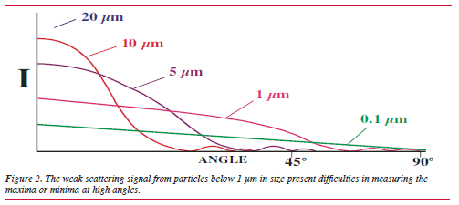
The technique has proven to be extremely accurate
for the sizing of both spherical and non-spherical
sub-micron particles.

Other manufacturers have begun to utilize
multi-frequency wavelength analysis for sub-micron
analysis, typically using just one additional wavelength,
though they do not adopt the same approach
as is taken for PIDS.™ While providing extra data, it
does not offer the same amount or level of detailed
information that is offered by PIDS technology. The
extra wavelength is normally provided by a blue
light source with a wavelength of approximately
460 nm. By shortening the wavelength, gains are
made by primarily ensuring a larger light flux signal
is generated compared to the standard laser light
source, thereby making its quantification easier.
Secondly, by using a shorter wavelength, the difference
between the small particles are minimalized, in
terms of size and the wavelength of the illuminated
light source; this is an important parameter for standard
diffraction analysis. Diffraction data, in effect,
stops being meaningful as particles get smaller in
relation to the wavelength that is irradiating them.

The Problem with Pigments

,p>Pigments provide a unique problem not encountered
with most materials that are measured using laser
diffraction instruments. The vast majority of samples
measured on commercially available instruments
are not colored and this makes their analysis more
straightforward. It is important to consider why this
is the case. For an accurate particle size to be calculated,
both the real refractive index of the material
and its imaginary component must be known. This
becomes more important for small particles as the
mathematical treatment to successfully size them
becomes the more rigorous Mie theory.

While the real refractive index is a value that is
well understood by the majority of analysts, the
imaginary component is less so. It is, in fact, the
degree of absorbance that is exhibited by the sample
at a given wavelength. Non-colored materials exhibit a
fairly uniform absorbence across the ultra violet/
visible (UV/Vis) electromagnetic spectrum.

Pigments, however, provide an entirely different
challenge. The reason they are colored lies with the
fact that they absorb certain wavelengths preferentially.
This must be taken into account when calculating
the particle size distribution, particularly if the
particles are small. For example, how will a blue
pigment with an absorbance maximum at 630 nm
interact with a helium neon laser (wavelength
633 nm), which is the choice of a number of manufacturers
for their primary laser light source? The material essentially behaves as a black body, which
must be taken into account during optical modeling.
Failure to do so will lead to significant errors that
will increase with a decrease in particle size.

The real refractive index remains an important
function and can be either calculated or estimated
from known constants, and any error associated
with this can be minimized.

Thus, it is easy to see that the quantification of
the imaginary component of the complex refractive
index is extremely important for the accurate determination
of pigment particulate systems.

Determining the Imaginary
Component

The determination of the imaginary component of a
pigment is a relatively straightforward measurement.
It is achieved using a UV/Vis spectrophotometer,
which measures the relative absorbency of a material
per given wavelength. It is this preferential absorbtion
that dictates the color of a material.

Two things must be taken into account: first,
one must ensure that no large particles are present
in the spectrophotometer sample cell, as they will
give rise to forward scatter and will “blind” the
detector, compromising the absorbance measurement.
It may be necessary to filter a sample to remove large
particles; particles should be no bigger than a few
microns.

Second, the relative amount of absorbtion must
be taken into account in the calculation of the optical
model. This will not be a constant and will need to
be determined for each type of instrument. Once this
has been done, these values can then be used when
calculating optical models for a given pigment.

In terms of the allowance used for the imaginary
component, the approach taken for each complementary
wavelength needs to be fully evaluated.
A novel approach related to the obscuration value,
which is associated directly with the amount of
sample in the analyzer, can be employed. A more
sophisticated method would be to use Beer’s Law.
However, one would need to know the exact concentration
of the solution. But, absorbencies taken
at a specific concentration could then be directly
related to one another. The more quantitative
approach would be to determine the exact concentration
of the solution.

Each manufacturer takes a different approach to
the calculation of optical models. Beckman Coulter
allows users to calculate a complete Mie theory optical
model for a given sample. The optical model can be
generated with the latest version of software in as
little as three seconds.

Using Complementary Information

When analyzing pigments, it is also beneficial to
utilize other sources of information to initially verify
or confirm the results obtained. Once correlating
information has proven the suitability of a given
model for a particular sample it can then be used
with confidence for that material. The best sources
of correlating information are photomicrographs.
These can be images from simple microscopes to
electron microscopes. This approach is particularly
important for the detection of small amounts of oversized
materials. This can be a common problem with
many pigment systems due to the type of size reduction
or milling techniques utilized. Ball mills can give
rise to small amounts of oversized materials that may
remain undetected by laser diffraction, particularly
if the imaginary component of the refractive index
is not taken into account.

Beckman Coulter, working in conjunction with
a selected number of pigment manufacturers, has now
gained valuable experience in the particle size
analysis of many different pigment systems. The
applicability of an optical model for a given pigment
is best determined if one tracks a milling process over
time. If the correct refractive index values have been
used to create the optical model, one should obtain
a constant reduction of the mean size.
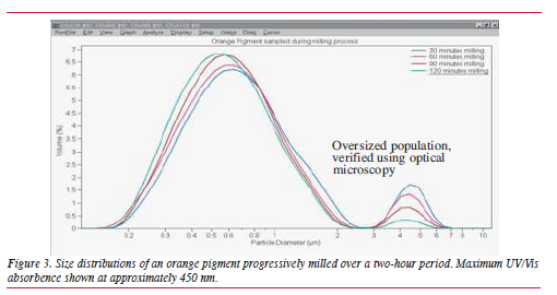
The Question of Shape

A criticism leveled at all laser-based particle sizing
devices is that they make no allowance for the shape
of the materials under test, regardless of the size of
the particles. The reasons for this lie with the underlying
assumptions, used in calculating size distributions
from the raw data generated during the analysis.

The mathematical models used to calculate distributions
are based on scattering of light by a
sphere. So any reported distribution is, in effect, an
equivalent spherical distribution of the material being
analyzed. In most instances this is quite adequate
since most particles approximate to a spherical system
adequately enough.

Particles in milled pigments will not be perfect
spheres, so how does this affect their recovered
sizes using the technique described above? The
ideal way to evaluate this is with reference to
known standards.

Until recently, sourcing suitable sub-micron,
non-spherical particles for studies of this type has
been difficult because of the lack of independently
produced and assayed materials. However, the colloid
chemistry department at the University of
Utrecht now produces a variety of mono-dispersed,
non-spherical materials. Figure 6 shows the results
obtained for the analysis of sub-micron hematite
spindles (spheroids).

The size of the particles (determined by SEM,
or scanning electron microscopy) is 46.9 nm in
width and 130.8 nm in length, giving them an
approximate aspect ratio of 3:1. The particles are
mono-sized and are readily dispersed.
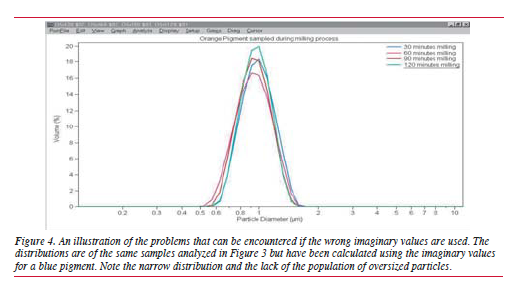
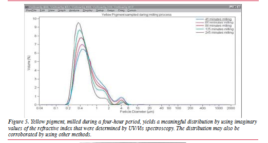
LS™ Series Particle Size

Distribution for Hematite spindles
The optical properties of the hematite spindles have
been determined from UV/Vis spectroscopy for the
imaginary component of the refractive index and
from UV/Vis spectroscopic ellipsometry data for the
real component. Using hematite has an added benefit,
in that being a colored material it mimics a pigmented
material well.

The reported value for the mean size from the
LS Series Analyzer is 78 nm (Figure 7), which is
well with in the range of what one would expect
given the random motion of the particles in the sample
cell. Statistically, one would expect the reported mean
size to be a function of all the possible orientations
of the particles as they traverse through the illuminated
beam. Indeed, instruments are designed to
ensure that particles in the sample cell are orientated
in a random manner with regard to their morphology
or shape.

Summary

If the right approach is taken, enhanced multi-frequency
laser diffraction can be employed successfully
to size particulate pigment systems.

For pigments, steps can be taken to determine
the imaginary component of the optical model for
laser diffraction particle size analyzer. It is also
worthwhile considering using other techniques to
initially corroborate the results obtained.
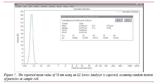
Continue reading

  • 1
  • 2

Have questions or need support?

1. Get in Touch
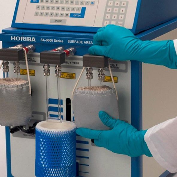
Tell us about your application and particle characterisation needs.

2. Have a conversation

We're happy to set up a call to discuss your application and answer your questions.

3. Discuss next steps

Expand your knowledge with a seminar, demonstration, sample analysis, or obtain a quote.

We are here in the UK to help and
answer any questions you might have.
Call us directly on +44 1582 704807

© 2024 Meritics.com